Clinical applications of advancing high frequency ultrasound techniques
By Dr Adrian Lim and Professor David Cosgrove, Department of Imaging, Imperial College London, United Kingdom
High frequency ultrasound (US) techniques continue to improve with better resolution and exquisite B-Mode imaging, particularly with improved compounding techniques seen with the Aplipure product.
However, particular focus has been on improving techniques for breast imaging and one distinctly novel idea is to highlight microcalcifications within tissues.
Micropure – This highlights those microcalcific foci as a ‘twinkling’ focus on a dark blue background (the latter can be altered to personal taste). This is the first time that an ultrasound device has been developed to help the operator confidently visualise microcalcifications, which previously would have been deemed too difficult with just conventional US. This could be particularly helpful with biopsy, where traditional methods would necessitate stereotactic sampling. The latter would require 10–20 core biopsies whereas, with the help of US guidance, this could be reduced, although its efficacy remains to be proven. Fig. 1 illustrates how the microcalcific foci in the 12 o’clock position of the right breast, seen mammographically, is more clearly seen with the MicroPure application.
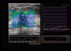
Elastography – The technique has been shown to distinguish benign from malignant breast lesions successfully. It is a method to quantify ‘manual palpation’ ultrasonically: the stiffer a lesion, the more likely it is malignant. Much of the published work has concentrated on providing colour maps of the area of stiffness of a lesion and surrounding tissues. Toshiba has added this capability to the Aplio, but provides an extra dimension by allowing time elasticity graphs to be plotted over a region of interest in the compression or relaxation cycles. By quantifying elasticity it removes the subjectivity of colour maps and some recent pilot studies (as yet unpublished) have suggested that the ratio of the adjacent normal fatty breast tissue to the lesion can be an indicator of malignancy when the ratio is at least 10. However, there are exceptions to the rule, particularly if a malignant lesion is necrotic or in cysts, which can also show stiffness. Fig. 2 displays the elastograph map and graph of a breast carcinoma.
Volume imaging – Another area of US development has been the ability to generate 3-D volumetric images with high frequency probes. Particularly successful in obstetrics, this has yet to find its clinical application in general imaging. However, it has become apparent that the coronal plane reformatted images of breast cancers provides an appreciation of the retraction of a lesion and perhaps gives a better estimation of the true size and extent of the mass. Further trials are needed to assess whether this really does provide a better representation of tumour size. Fig. 3 shows the extent of a carcinoma with the ‘finger like’ projections closely resembling those seen on the MRI images. 3-D elastography techniques have also been suggested to be helpful in assessing breast mass lesions.
Contrast enhanced ultrasound – Although US contrast agents have an established use in abdominal studies, particularly for characterising focal liver lesions, they have
yet to find their niche with superficial lesions and high frequency scanning, particularly for breast lesions. Fig. 4 shows how the enhancement pattern and angiogenic vessels of a malignant tumour can be depicted with microbubble enhancement. It remains controversial as to how much this adds for distinguishing benign from malignant lesions, since breast biopsies are easily performed with few complications or significant morbidity. The role of contrast enhancement may therefore not be major; however, it may find a role in assessing response to treatment.
Conclusion – There is immense potential for the latest developments in breast ultrasonography, in particular Micropure and elastography time/stiffness curves, which provides quantification and does not rely solely on subjective visualisation. Together with the 4-D and small parts contrast capabilities, the new version 3 Aplio XG provides the radiologist with a great armamentarium for evaluating complex breast lesions.
It is not envisaged that Micropure would replace mammography in screening for microcalcifications, but the ability to biopsy microcalcifications under US guidance, and thus obviating the need for stereotactic biopsies, would be of notable value. The ability to depict the true extent of malignant breast tumours more accurately with elastography, 4-D imaging and the use of microbubbles would not only help surgical management but also has potential in assessing response to chemotherapeutic treatment, thereby aiding the oncologist.
The potential of these techniques to detect problematic breast tumours such as lobular or multifocal breast carcinomas, as well as in screening, should be fully investigated. Ultimately, these latest developments require multicentre studies to evaluate their true value and potential.
28.10.2008