Article • CEUS
Only convincing with contrast
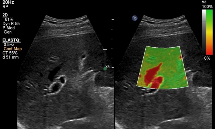
Contrast enhanced ultrasound is part of clinical routine and makes a substantial contribution towards increasing the significance of tissue and organ diagnosis with ultrasound for difficult cases.
‘CEUS allows us to optimise our daily routines, to achieve the correct diagnosis faster and to improve solutions for unclear cases,’ explains Professor Dirk-André Clevert MD, head of the Interdisciplinary Ultrasound Centre at the Ludwig Maximilian University of Munich. The emergence of new competitors in the market and increasing competition between providers further enhances technological innovations.
Ultrasound, and specifically contrast ultrasound, is not inferior to cross-sectional imaging. Particularly in cases where the result of a CT or MRI scan is not clear, ultrasound often provides an important second opinion. Unlike a primary radiological diagnosis contrast ultrasound does not just deliver a snapshot but an entire image or, as Clevert puts it, the entire news programme, from the welcome to the weather forecast. ‘The advantage of ultrasound is its high temporal resolution, which really allows us to observe how the microbubbles flow over a period of several minutes. We can see the process and also observe what happens afterwards, which is especially helpful in cases where the process is not quite typical.’
In his opinion, the comparison between cross-sectional imaging and ultrasound is also impaired because CT or MRI scanning with contrast media is often compared to native ultrasound. Comparing both technologies without contrast media would be much closer to the truth and reality.
For Clevert it is therefore logical that CEUS is now licensed in its primary field of application for liver diagnostics, and specifically to detect and characterise liver lesions, in the USA, Canada and Brazil, with Mexico soon to follow this example.
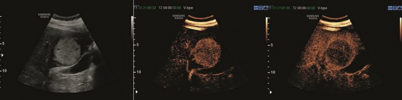
Clevert has also experienced the benefits of off-label use of CEUS, especially for children. The use of contrast-enhanced ultrasound in children is governed by very strict rules. An example of how helpful it can be is the case of a young girl with an aortic isthmus stenosis. The two-and-a-half-year-old had been fitted with an aortic stent, but suffered a partial tear of the external iliac artery when the sheath was withdrawn. After surgery, using a patch, a large haematoma developed in the abdomen which was to be punctured with the help of ultrasound, because the CT scan showed no bleeding. Only the contrast ultrasound image actually showed a small leak in the patch.
Consequently, no puncture was carried out, but immediate surgery to remove the haematoma and revise the patch. ‘Although the use of contrast medium in this case was off-label,’ says Clevert, ‘the medical team were within their rights to utilise CEUS, as its use paid off for the patient 100%.’
Not all problems can be solved by using ultrasound, however. As a rule, the following applies: The better an organ can be visualised with ultrasound the better the results of contrast-enhanced ultrasound will be. If visualising the liver is difficult because of meteorism contrast-enhanced ultrasound also cannot rule out liver lesions. ‘The better the basic settings, the cleaner the use of ultrasound, the better the result achieved with CEUS. Obviously you also need a good system for this,’ he points out. ‘We can’t expect the same performance from a mid-range scanner as from a high-end system. In other words, we have to adapt our expectations to the equipment!’
There are some very exciting questions for ultrasound and there is still a way to go to fulfil all the demands from high-end users.
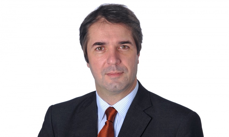
Prof. Dirk-André Clevert
Clevert is pleased that Samsung has now penetrated the established market for high-end equipment. Most companies have decades of experience and can adapt their systems to customer specifications. ‘If you want to compete in this market your system must have an edge over those offered by the competition. As a new player in this market, Samsung had to make large investments to get to the point where a scanner is not just a ‘nice to have tool’ but diagnostically innovative. But the company is gaining ground, aided by its fast development of software and the openness towards suggestions from clinicians,’ Clevert enthuses.
Within clinical routine, Samsung’s RS80A with Prestige mostly wins over in workflow. The company is at an advantage here, due to its extensive experience with monitors in the consumer sector. The touchscreen is of a particularly large, convenient size and clearly arranged. An additional, flexible, portable touchscreen, which could be used to present the day’s caseload during meetings, would be beneficial, but would basically be a ‘nice to have tool.’
What is really essential for high-end equipment is good, contrast enhanced ultrasound, whilst compression and shear wave elastography are now also a ‘must’, and so is image fusion, and the RS80A with Prestige offers all of this. ‘There are some very exciting questions for ultrasound and there is still a way to go to fulfil all the demands from high-end users,’ Clevert concludes. ‘But Samsung is definitely heading in the right direction.’
Profile:
Berlin-born Professor Dirk-André Clevert began his career at the MRI Diagnostics Institute Westend in that city and in the Department for Internal Medicine at the Waldkrankenhaus Gransee. He then worked as a specialist registrar in the Department of Radiology at Passau Hospital for three years. In 2003, he moved to Munich and joined the newly founded (2004) Interdisciplinary Centre for Ultrasound at the Grosshadern Campus, University Hospital Munich. The centre coordinates all ultrasound activities in the hospital. As course director and congress president, he organises numerous national and international ultrasound courses and congresses. On the 80th founding anniversary of the Medical Faculty of Tbilisi State Medical University, Clevert, as head of the Interdisciplinary Ultrasound Centre, was awarded an honorary doctorate.
25.10.2016