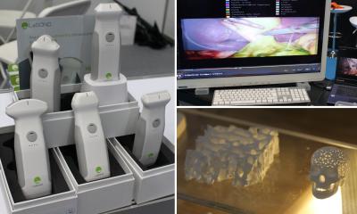
Portable real-time 3-D ultrasound for brain scans
USA - 3-D ultrasound technology developed at Duke University provides images of the brain vessels in real-time, which could be miniaturised in coming years for use in ambulances. In an initial pilot study, the system has passed the proof-of-principle.
The researchers used a matrix phased-array transducer to scan a 65-90 pyramid, producing up to 30 volumetric scans per second. A way was found to compensate for different skull thicknesses, which, if uncorrected, might result in poor resolution and contrast. The ultrasound transducer must be held motionless for 20 seconds while data on the echoes passing through bone are processed. The team predicts that future systems should reduce the required time to under a second.
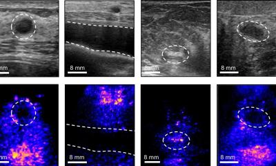
Doppler imaging was performed before and after administering a microbubble contrast agent to 17 healthy volunteers. Their heads were scanned with the ultrasound transducer placed on each temple. Nine more volunteers were examined from the neck base up.
A vascular sonographer and a neurologist reviewed the images
for independent evaluations. They detected the vessels on the side in 71% of the cases and the complete vessel circle in 51%. They also identified the entire vertebrobasilar circulation in 22% and the basilar artery in 44% of the nine volunteers.
To determine the diagnostic value for transcranial ultrasound, the team now plans to test the image correction technique in more volunteers. Additional studies are envisaged to asses the sensitivity and specifity
of contrast-enhanced, real-time 3-D transcranial ultrasound compared with either digital subtraction angiography or MR angiography.
19.11.2008