Microbubbles in tumour therapy
Contrast agents have opened up entirely new possibilities are taking shape for ultrasound, above all in oncology. Following the publication of guidelines on the clinical use of contrast-enhanced ultrasound (CEUS) by the European Federation of Societies for Ultrasound in Medicine and Biology (EFSUMB) in 2004 and 2008, at this year’s World Ultrasound Congress, WFUMB and EFSUMB will present joint CEUS guidelines for the liver and other organs. Professor Gerhard Mostbeck MD, the WFUMB Congress President and Chairman of the Organising Committee, explains the promises CEUS holds for tumour therapy.
‘The development of the second generation contrast agents in the past four years has made CEUS much easier in clinical practice,’ said Professor Mostbeck. ‘In turn, this meant that the procedure has been approved for many more indications. Also, due to developments on the medical technology side, CEUS is now more widely available. Today, many ultrasound systems are equipped with dedicated software programmes tailored to contrast-enhanced visualisation.
‘Unfortunately, in many countries, such as Austria, the medical insurers do not reimburse the costs of contrast agents. Consequently CEUS is rarely performed. Office-based radiologists, for example, generally don’t offer this procedure because it’s not covered by insurance. We definitely need to convince the payers to support CEUS. We have already been able to prove that the use of contrast-enhanced ultrasound is indeed cost-efficient and helps reduce the number of CT and MRI exams.
How can CEUS realise new cancer treatments?
‘In oncology, an objective assessment of the tumour response to a certain treatment provides relevant information on the therapy effect, as well as in a research context. To date, diagnostic imaging has been used primarily to gather data on tumour size. In this respect ultrasound is not the ideal modality, because many studies require tumour size measurement before and after the therapy – something ultrasound is unable to deliver because ultrasound images are difficult to reproduce.
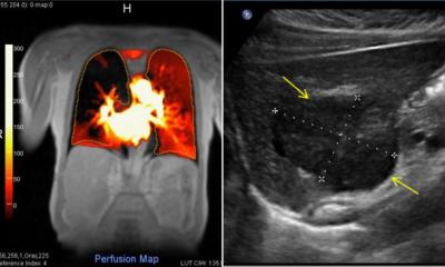
‘Modern anti-cancer drugs aim to inhibit tumour angiogenesis. Effective therapy does not necessarily mean that the tumour shrinks; much rather, vascular density in the tumour tissue is reduced. The use of contrast agents offers a new possibility to assess tumour perfusion. Since anti-angiogenetic tumour treatments can be very expensive, it makes sense to gather information on the therapy effectiveness as early as possible. Some current studies on this issue will be presented at the congress.
When are contrast agents particularly helpful?
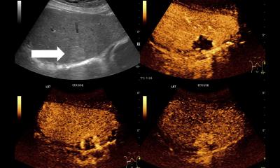
‘Contrast-enhanced ultrasound is indicated particularly with well visible lesions, as in breast cancer, liver metastases or renal cell carcinoma. However, we don’t have sufficient data yet. It will take another few years until the Response Evaluation Criteria In Solid Tumours (RECIST) in CT and MRI can be replaced.
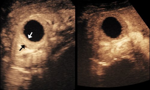
‘Nonetheless, I consider CEUS to be an important step in the right direction in order to develop quantitative parameters on tumour perfusion. We tried colour Doppler, but since the assessment of tumour vascularity was not really quantifiable, this method – quite justifiably – has never reached clinical practice. But now there is a seriously promising method to assess tumours sonographically, right after completion of the initial therapy cycle and to store the information as data that can be easily reproduced. The method is not yet in phase three or four of clinical studies but the results so far have been very encouraging.
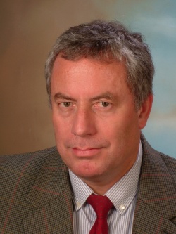
Gerhard Mostbeck
Professor Gerhard Mostbeck MD received specialist training and earned his professorship with a thesis on duplex sonography of the renal vessels, at the Central Institute for Radio Diagnostics at the University of Vienna.
The radiologist was inter alia President of the Austrian Society of Ultrasound in Medicine (2002-2005), of the Austrian Röntgen Society (2006-2008) and of the Joint Meeting of the Austrian, German and Swiss Societies for Ultrasound in Medicine in 2000.
Today, he is Director of the Institute for Diagnostic and Interventional Radiology and Radiological Outpatient Services at Vienna’s Wilhelminenspital, and Director of the Institute for X-Ray Diagnostics at the Social Medicine Centre Baumgartner Höhe Otto Wagner Spital, Vienna.
25.08.2011