© Roberto Schirdewahn
News • High-resolution imaging
Ultrasound reveals vascular details with microbubbles
A touch of the button is all it takes to improve the resolution of ultrasound images fivefold. This is down to bubbles floating in the bloodstream and a clever algorithm.
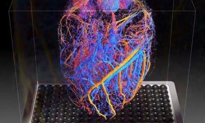
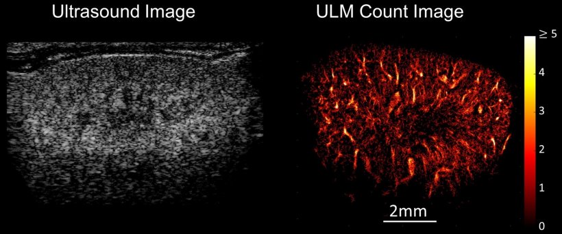
The tiniest vascular structures have remained elusive for ultrasound imaging – until now. A research team from the Department of Medical Engineering at Ruhr University Bochum headed by Professor Georg Schmitz is perfecting ultrasound localization microscopy (ULM). By deploying a commercially available contrast agent with microbubbles and performing a number of calculation steps, it’s now possible, for example, to image the vascular structure of a mouse kidney in minute detail, as well as the vascular structure of tumors.
© Roberto Schirdewahn
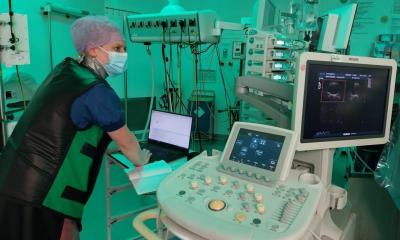
Schmitz and his team have been refining this technique for over ten years. In 2011, they came up with the brilliant idea of using a contrast agent consisting of microbubbles for the high-resolution imaging of vessels in ultrasound. They follow the flow of blood into the smallest vessels and reflect ultrasound waves so well that they light up white in the image. “The contrast agent only remains in the body for around ten minutes, then the bubbles are broken down and the gas is exhaled through the lungs,” explains Georg Schmitz. It only takes 30 to 90 seconds to take the images.
In order to increase the resolution of an ultrasound image so dramatically, it’s necessary to perform several intermediate steps. The involuntary movements of the patient’s body must be taken into account. In the next step, the algorithm developed by the researchers removes the background from the images so that only the vesicles are displayed. Then, the center of each individual bubble must be marked. And finally: the tricky calculation of which bubble has traveled along which path. “Our algorithm always looks at a group of neighboring images and uses the highest probability to decide which path a bubble has taken,” explains Georg Schmitz.
© Georg Schmitz
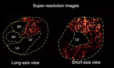
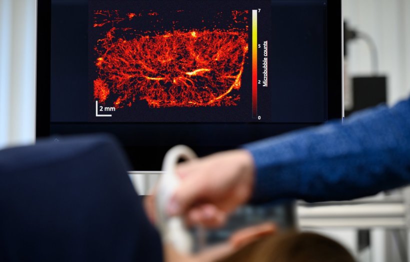
What ultimately emerges from the entire calculation after a few minutes is an image of the system of small vessels through which the bubbles have moved. It’s even possible to read off the direction and speed in which the bubbles have streamed through.
It allows [physicians] to identify the characteristics of a tumor, for example: Where is it supplied from? What do the supplying blood vessels look like?
Georg Schmitz
“The direction of movement and the shape of the small vessels through which the blood flows are information that physicians need to know precisely,” stresses Georg Schmitz, who has worked with clinical partners on several projects. “This is because it allows them to identify the characteristics of a tumor, for example: Where is it supplied from? What do the supplying blood vessels look like?” Such information can provide insight into how aggressive a disease is and possibly also which therapy will or will not work. Looking at the smallest vessels is also valuable for monitoring the effect of chemotherapy. Ultrasound localization microscopy is currently the only clinical imaging method that can make such fine blood vessels visible at a depth of several centimeters.
Source: Ruhr University Bochum
04.06.2024