Application of Vascular Recognition Imaging (VRI)
Vascular Recognition Imaging (VRI) is a low MI broadband colour Doppler method that images the interactions of Sonovue with microbubbles in a non-destructive manner as perfusion images of a quality never before achieved. The perfusion image represents the low velocities of unmoving or barely moving microbubbles as green-coded tissue perfusion, while the higher velocities of the veins and arteries are imaged according to convention in red or blue coding.
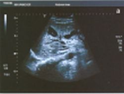
Fig. 7: 30-year-old patient who developed several liquid areas in the right liver lobe after falling off a horse (a).
This article was first published in the VISIONS, issue 4/2003, a publication of Toshiba Medical Systems
17.08.2007
More on the subject:More on companies: