News • Ophthalmology
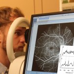
Portable AI-powered scanner to facilitate access to eye care
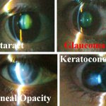
Researchers are developing a portable AI-powered scanning slit-light device, designed to make ophthalmic care more accessible, so patients can be assessed any place, and any time.