A soft tissue probe for in vivo imaging of gynaecological tissue
A probe suitable for imaging soft tissue for use with the VivoSight Multi-Beam optical coherence tomography (OCT) imaging system has been launched by manufacturer Michelson Diagnostics. The probe enables in vivo imaging of oral and
gynaecological tissue.
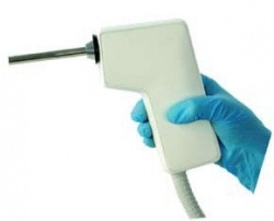
‘The VivoSight Multi-Beam OCT system provides sub-surface cross-sectional images at far higher resolution than is possible with ultrasound, CT or MRI, and much deeper and wider than is possible with confocal microscopy,’ Michelson reports. ‘The new Soft Tissue Probe provides the same unprecedented imaging quality as the Topical Probe, with real time, in vivo images at better than 7.5 μm lateral resolution. The probe is 9 cm long and provides both 2-D and 3-D images over a 5 mm x 5 mm area. For sterile applications, a disposable transparent sheath covers the probe, handle and upper connecting cable.
‘The structures of the epithelial layers are exquisitely revealed in amazing detail,’ said the firm’s CEO Jon Holmes. ‘The new probe promises to be a tremendous aid to clinicians for the diagnosis and treatment of oral and cervical cancers.’ Ex vivo trials on excised oral tissue have shown that Multi- Beam OCT can visualize structures such as the epidermal / dermal junction and areas of cellular crowding characteristic of early stage tumours. Michelson’s clinical partners have reported achieving sensitivity of 80% and specificity of 81% for oral cancer diagnosis, from a blinded assessment of OCT images of 125 excised oral lesions.
It is expected that in vivo imaging will give even better results and could eliminate the need for a biopsy, the firm adds. ‘The probe will also be suitable for imaging tumour margins prior to surgery.’
07.07.2010