First OCT tissue images
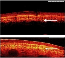
A UK collaboration, led by an optical imaging company has produced its first optical coherence tomography (OCT) tissue images at a wavelength of 1 µm. Based on interferometry, OCT works by analysing near-infrared light reflected from a sample and using differences in the reflected intensities to create high-resolution images. Employing 1 µm light - as opposed to the longer wavelength of 1.3 µm that's more commonly used for imaging skin - can ramp the quality of the resulting images.

"We believe that images acquired at 1 µm will offer improved contrast and resolution that will help clinicians to distinguish between healthy and cancerous tissue," said OCT expert Wolfgang Drexler, director of research at the University of Cardiff School of Optometry and Vision Science.
Initial images of healthy skin tissue, obtained using a Michelson Diagnostics OCT microscope converted for 1 µm operation, appear to support this view. Features such as capillaries and the boundaries between tissue layers are more clearly defined than in a comparable 1.3 µm OCT scan.
The project, entitled OMICRON, is developing an in-vivo OCT imaging probe - operating at the new wavelength of 1 µm - for high-resolution sub-surface tissue imaging (see The optical biopsy: coming into view?). The long-term objective is to use such a system to perform real-time diagnosis and guide the removal of cancerous or precancerous tissue during the same procedure.
Currently, clinicians diagnosing, treating and monitoring cancer frequently take biopsy samples of tissue for analysis by pathologists. This process can be inefficient, expensive and time consuming. In addition, the biopsy may be poorly targeted on the tumour, and results from the laboratory analysis can take weeks to come back.
"OCT scanning has the exciting potential of both guiding and reducing the dependence on biopsies," explained Nick Stone, head of biophotonics at Gloucestershire Hospitals NHS Foundation Trust. "This could speed up cancer diagnosis and treatment, reducing the pressure on overloaded pathology departments, and improve outcomes from cancer surgery."
Michelson Diagnostics says that further work in 2009 will concentrate on boosting the laser power to increase the depth penetration, as well as evaluating and analysing images of cancerous tissue taken with the new probe.
• OMICRON was funded in 2007 by the UK's Technology Strategy Board. Partners in the collaboration are: Michelson Diagnostics; the University of Cardiff; Gloucestershire Hospitals NHS Foundation Trust; the National Physical Laboratory; semiconductor specialist Kamelian; and medical imaging systems specialist Tactiq.
This article was first publish at www.medicalphysicsweb.com
11.11.2008


