News • Biological image analysis
Machine learning accelerates super-resolution microscopy
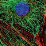
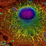
Scientists use super-resolution microscopy to study previously undiscovered cellular worlds, revealing nanometer-scale details inside cells. This method revolutionized light microscopy and earned its inventors the 2014 Nobel Prize in Chemistry. In an international collaboration, AI researchers from Tübingen have now developed an algorithm that significantly accelerates this technology.