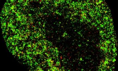
Image source: Adobe Stock/Dr_Microbe
News • New high-tech microscope
Using AI to detect malaria in returning travelers
An automated microscope combined with AI software can identify malaria parasites in blood samples almost as accurately as experts staffing microscopes used in standard diagnostic procedures, according to research at University College London Hospitals (UCLH).
Use of the system as an additional diagnostic approach to disease detection may help reduce the burden on microscopists, the research found. Malaria is an infectious disease claiming more than half a million lives each year. Because traditional diagnosis takes expertise and the workload is high, an international team of researchers investigated if diagnosis using a new system combining an automatic scanning microscope and AI is feasible in clinical settings.
Writing in Frontiers in Malaria, the international team of researchers assessed whether a fully automated system, combining AI detection software and an automated microscope, can diagnose malaria with clinically useful accuracy.
“At an 88% diagnostic accuracy rate relative to microscopists, the AI system identified malaria parasites almost, though not quite, as well as experts,” said Dr Roxanne Rees-Channer, a researcher at The Hospital for Tropical Diseases at UCLH, where the study was performed. This level of performance in a clinical setting is a major achievement for AI algorithms targeting malaria. It indicates that the system can indeed be a clinically useful tool for malaria diagnosis in appropriate settings.”
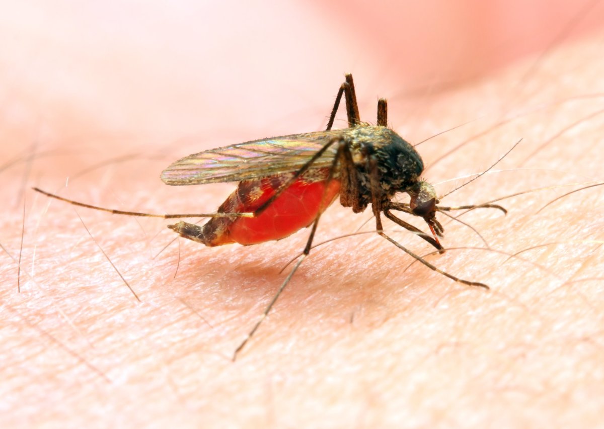
Image source: Adobe Stock/Kletr
The researchers sampled more than 1,200 blood samples of travelers who had returned to the UK from malaria-endemic countries. The study tested the accuracy of the AI and automated microscope system in a true clinical setting under ideal conditions. They evaluated samples using both manual light microscopy and the AI-microscope system. By hand, 113 samples were diagnosed as malaria parasite positive, whereas the AI-system correctly identified 99 samples as positive, which corresponds to an 88% accuracy rate.
The fully automated malaria diagnostic system the researchers put to the test includes hard- as well as software. An automated microscopy platform scans blood films and malaria detection algorithms process the image to detect parasites and the quantity present.
Automated malaria diagnosis has several potential benefits, the scientists pointed out. “Even expert microscopists can become fatigued and make mistakes, especially under a heavy workload,” Rees-Channer explained. “Automated diagnosis of malaria using AI could reduce this burden for microscopists and thus increase the feasible patient load.” Furthermore, these systems deliver reproducible results and can be widely deployed, the scientists wrote.
Despite the 88% accuracy rate, the automated system also falsely identified 122 samples as positive, which can lead to patients receiving unnecessary anti-malarial drugs. “The AI software is still not as accurate as an expert microscopist. This study represents a promising datapoint rather than a decisive proof of fitness,” Rees-Channer concluded.
Source: University College London Hospitals
15.08.2023