News • Mitosis profiling
AI tool helps grade cancer based on cell divisions
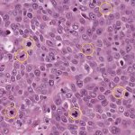
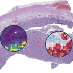
Ahead of World Cancer Day on 4 February, scientists are revealing a cutting-edge artificial intelligence (AI) tool designed to help grade cancer, by analysing cell division.


Ahead of World Cancer Day on 4 February, scientists are revealing a cutting-edge artificial intelligence (AI) tool designed to help grade cancer, by analysing cell division.
The underlying mechanisms of Guillain-Barré Syndrome (GBS) have remained largely unknown until now. New research now uncovers a pivotal aspect of GBS pathophysiology.

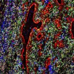
Bringing digital pathology together with novel multiplexed staining techniques may answer key questions about complex diseases. Pathologist Lukas Marcelis, MD, PhD, believes such combinations of technology will have benefits for clinicians and patients and can help unravel some of the mysteries surrounding a range of conditions.

White blood cells found in breast tumors can both help and hinder the spread of cancer cells to other organs, a new study from Karolinska Institutet shows.

Breaking & entering – on a minuscule scale: US researchers have shown how some pathogens like Toxoplasma can enter cells using physical force to cause an infection.
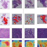
Research from Shenzhen proposes an integrated diagnosis model for automatic classification of adult-type diffuse gliomas directly from annotation-free standard whole-slide pathological images.

Cedars-Sinai Medical Center, a non-profit hospital and medical research institution in Los Angeles, is setting new standards for quality and innovation in patient care by successfully introducing typing of Candida auris species – a procedure that could prove crucial in protecting patients from infection outbreaks caused by these microbes in healthcare settings.
Researchers from Finland have developed an artificial intelligence tool for automatic colorectal cancer tissue analysis that outperforms prior methods.
For the first time, researchers show that AI-based predictions can deliver comparable results to clinical tests on biopsies of patients with colorectal cancer (CRC).

Experts have highlighted how precision pathology using Artificial Intelligence can provide an effective alternative to molecular diagnostics. This, say a team from the Karolinska Institutet (KI) in Stockholm, Sweden, can also offer multiple advantages within a clinical setting and support risk stratification.
An 'encyclopaedia' of protein alterations in soft tissue sarcomas could open the door to a new era of understanding and treatment for this group of rare cancers.

A new study led by researchers from the University of Notre Dame links a high body mass index (BMI) to alterations in the structure and environment of cancerous tumors.

With the introduction of digital pathology, the University of Queensland and Sullivan Nicolaides Pathology aim to provide significantly improved tests in terms of cost, quality and speed.
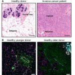
Researchers have found a possible explanation as to why higher breast density and older age increase the risk of breast cancer. According to the experts, adipocytes play a vital role here.
A research team at UCLA has made an important advancement to address one of the major challenges in cell-free DNA (cfDNA) testing, also known as liquid biopsy.

From current mixed reality assistance, technological advances could soon make fully digital autopsies a reality. This could enhance efficiency and legal certainty, and also benefit training.

Researchers at Harvard Medical School developed a new tool that promises to improve the way pathologists see and evaluate a tumor by providing detailed clues about the cancer.
A study led by the National Institutes of Health’s RECOVER Initiative and supported by NYU Langone Health provides an expanded working definition of long Covid.

Lyme disease, the most common tick-transmitted bacterial infection in the world, is challenging to diagnose. Initial early-stage symptoms may include skin rash, fever, headache, fatigue, swollen lymph nodes, and/or body and joint aches. However, these symptoms are also associated with many other diseases and medical conditions.
It is crucial that labs can rely on their slides for a seamless – and accurate – diagnosis. With many more commercial instruments focusing on flexibility and choice, these qualities are difficult to separate from subjectivity and complexity.

The Netherlands Cancer Institute (NKI) will deploy a diagnostic platform from digital and computational pathology solutions provider Proscia, the company announced.

A new blood test that can track and follow the neurodegeneration in Alzheimer's disease - and exclude other dementias.
Dutch scientists have discovered how specialized immune cells can detect and remove cancers that are ‘invisible’ to the conventional defense mechanisms of the immune system.
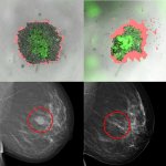
Swedish researchers have developed a method that should be able to predict whether a patient with breast cancer will benefit from a particular treatment or not.

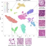
New research shows the power of artificial intelligence (AI) applied to endometrial carcinoma microscopy images. This could improve diagnosis and treatment of uterine cancer.