News • Towards personalised risk assessment
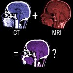
Training an AI to early detect dementia with big data
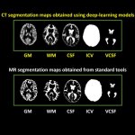
With enough medical training data, AI can predict health conditions with astounding accuracy. Now, researchers want to use brain scans of the entire Scottish population to have an algorithm early detect dementia.