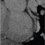
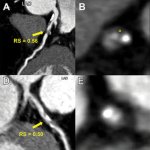
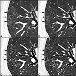
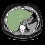
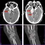
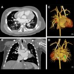
CT

Photo

News • Radiology research

Chest CT illuminates COPD mortality risk

Body composition information derived from routine chest CTs can provide important information on the overall health of people with chronic obstructive pulmonary disease (COPD), including their risk of all-cause mortality. This is the result of a new study, published in Radiology. COPD is a group of chronic, progressive lung diseases that affect about 30 million people in the United States alone.…

Photo

Article • Liquid metal bearing technology

Keeping CT scanning running smoothly

Liquid metal bearing (LMB) technology has been a key aspect of extending the lifespan of CT scanners, improving workflow, and enhancing patient and operator experience via noise reduction. The LMB concept has been pioneered by Dunlee, with senior CT product manager Robert Bayerlein noting that the Hamburg-based company has a long heritage in imaging innovation.

Photo

Sponsored • Radiology collaboration

Improved workflow and a touch of Disney magic

Improving workflow is one of the major challenges that radiology departments face. The need to be more efficient, deliver timely and effective patient care, and keep an eye on costs are all factors that seem to be ever-present in the modern imaging department. With the added demands of the coronavirus pandemic as radiology departments continue to play a critical role in the fight against…

Photo

Sponsored • All-in-one imaging trailer

Mobile solutions for the COVID-19 frontline

With public health issues continuing to make daily mainstream news headlines across the world, it is clear how much change the healthcare environment is going through. Not only are there existing pressures on resources, space, staff, certain procedures, and budgets, but there are brand new ones resulting from the ongoing global COVID-19 pandemic. This has led to an even greater need for…

Photo

News • Image-based diagnosis of Covid-19

AI detects coronavirus on CT scans

In order to detect the Corona virus SARS-CoV-2, there are further methods of diagnosis apart from the globally used PCR tests (Polymerase chain reaction): The infection can also be recognised on CT scans – for which Artificial Intelligence (AI) can be used as well. An AI system can not only filter CT scan of Covid-19 patients from a data set, but also estimate, which areas of the image are of…

Photo

News • Fight against COVID-19

AI lung scan analysis rolled out across Europe

The Belgian initiative icovid, which supports radiologists in the assessment of CT images of the lungs of COVID-19 patients, has grown into a multicentre European project, co-funded by the EU Horizon 2020 programme. Icovid was set up in March by UZ Brussel, KU Leuven, icometrix and ETRO, an imec research group of VUB.

Photo

News • CT & ultrasound fusion

‘Virtual biopsies’ could replace tissue biopsies in the future

A new advanced computing technique using routine medical scans to enable doctors to take fewer, more accurate tumour biopsies, has been developed by cancer researchers at the University of Cambridge. This is an important step towards precision tissue sampling for cancer patients to help select the best treatment. In future the technique could even replace clinical biopsies with ‘virtual…

Photo

Sponsored • Eliminating the need for additional monitors

New displays for different diagnostic images

Displaying medical images from different modalities such as CT, CR/DR, MR, ultrasound or mammography and pathology on one monitor - that is what most radiologists want. Many of them still use several medical displays next to each other. JVCKenwood's new CL-S1200 30.9-inch colour monitor makes this a thing of the past. The 12 megapixel device (4,200 horizontal and 2,800 vertical) can display…

Photo

Article • Coronavirus imaging

Covid-19: Is CT more sensitive than PCR testing?

Covid-19 causes characteristic changes in lung tissue visible in CT scans and chest radiographs, known as “ground-glass” opacities. Imaging is now considered a valid alternative, possibly even superior to RT-PCR. ‘This sparked an international debate about the role of CT in the diagnostic work-up of Covid-19,’ said radiologist Professor Cornelia Schäfer-Prokop.

Photo

Article • The role of chest CT in diagnosis and treatment

UPDATE: Covid-19 and lung infections imaging

RSNA 2020: International experts showcased new studies on chest CT’s role in Covid-19 diagnosis and treatment. A staggering volume of work and has been produced on the pandemic this year, with an average 367 Covid-19 journal articles published per week, according to Michael Chung, Assistant professor of radiology at Icahn School of Medicine at Mount Sinai, NYC.

Photo

Article • Modality comparison

COVID-19 imaging: lung ultrasound vs chest CT

A recent preprint study in France underpins the debate on whether lung ultrasound (LUS) should be used to triage COVID-19 patients better at the hospital as well as in primary care. The eChoVid study, published as a preliminary report of work on medTrix, shows that LUS enables identification of lung lesions as well as chest CT in COVID-19 patients. A team of French researchers compared routinely…

Photo

News • Improved accuracy and efficiency

AI could improve CT screening for COVID-19

Researchers at the University of Notre Dame are developing a new technique using artificial intelligence (AI) that would improve CT screening to more quickly identify patients with the coronavirus. The new technique will reduce the burden on the radiologists tasked with screening each image. Testing challenges have led to an influx of patients hospitalized with COVID-19 requiring CT scans which…

Photo

Video • Improving first aid

New 3D CT scanning method shows what happens during CPR

Rapid first aid during cardiac arrest makes the difference between life and death. But what happens to the heart and the internal organs when people come running and begin to give well-meaning but heavy-handed heart massage as they attempt to keep the person who has suffered a cardiac arrest alive? A research collaboration between the Department of Forensic Medicine at Aarhus University and the…

Photo

News • Corona in radiology

Revealing COVID-19-related brain injury in MRI and CT imaging

Injuries in the nervous system of patients with severe COVID-19 are revealed by magnetic resonance imaging (MRI) and computed tomography (CT). In a study of 185 patients, researchers at Karolinska Institutet (KI) and Karolinska University Hospital show an affection of microscopic blood vessels and inflammation in the brain, meninges and nerves. The results are published in Radiology.

Photo

News • Nanoparticles

Luminescent substance allows accurate viewing of body parts

Researchers have designed a fluid that works like a luminous ink to obtain very sharp images of damaged tissues, organs and cartilages in diagnostic tests. This new compound, still in the laboratory phase, reduces adverse effects on the human body because it allows lower amounts to be injected and the dose to be targeted only at the affected area.

Photo

Sponsored • Deep Learning in Radiology

New Levels of Precision with Self-learning Imaging Software

The complex form of machine learning DLIR (Deep Learning Image Reconstruction) is based on a deep neuronal network which is similar to the human brain. The artificial neurons of this network learn according to their biological model through intensive training. For the DLIR image reconstruction, the network is fed with sample data from phantom images on the one hand and high-resolution images of…

Photo

Article • Preparing for the unpredictable

The role of radiology in mass casualty incidents

CT has a critical role to play in management of mass casualty incidents with the ability to image patients from head to toe, offering a rapid overview for clinicians. The benefits of CT were outlined by Dr Elizabeth Dick during an ECR session examining the role of radiology in the management of mass casualty incidents, terror attacks and assaults.

Photo

Article • Goodbye to grayscale

Dual Energy CT – seeing x-rays with colors

Computed tomography (CT) is one of the most important imaging options for the diagnostics of patients. For a long time, in clinical routine CT technology mostly showed the Hounsfield Units (HU) that are indicated in the grayscale. However, the Dual Energy CT (DECT) enables colored X-ray images, therefore significantly improving examination methods. “We can also do material separation and get a…

Photo

News • Increased usability and precision

New X-ray contrast agent enhances vascular imaging

Researchers at the University of Zurich have developed a new X-ray contrast agent which is easier to use and distributes into all blood vessels more reliably, increasing the precision of vascular imaging. This reduces the number of animals required in research experiments. Various diseases in humans and animals – such as tumors, strokes or chronic kidney disease – damage the blood vessels.…

Photo

News • Inner ear disorder

Using advanced X-ray technology to fight Ménière's disease

The organ of balance in the inner ear is surrounded by the hardest bone in the body. Using synchrotron X-rays, researchers at Uppsala University have discovered a drainage system that may be assumed to play a major role in the onset of Ménière's disease, a common and troublesome disorder. These results are published in the journal Scientific Reports. Ménière's disease is manifested in sudden…

Photo

News • Algorithm-assisted diagnostics

AI in imaging: not as reliable as you'd think

Machine learning and AI are highly unstable in medical image reconstruction, and may lead to false positives and false negatives, a new study suggests. A team of researchers, led by the University of Cambridge and Simon Fraser University, designed a series of tests for medical image reconstruction algorithms based on AI and deep learning, and found that these techniques result in myriad…

Photo

News • Better tissue discrimination, lower radioation dose

Improving image quality of CT scans

Computed tomography (CT) is one of the most effective medical tests for analysing the effects of many illnesses, including COVID-19, on the lungs. An international team led by the Universitat Oberta de Catalunya (UOC) has developed a new method that improves the quality of the images obtained from CT scans. The algorithm, which has been tested on simulated data, enables them to distinguish…

Photo

News • What radiologists need to know

How lung disorders like COVID-19 affect children

Although the clinical symptoms of new pediatric lung disorders such as severe acute respiratory syndrome (SARS), swine-origin influenza A (H1N1), Middle East respiratory syndrome (MERS), e-cigarette or vaping product use-associated lung injury (EVALI), and coronavirus disease (COVID-19) pneumonia may be nonspecific, some characteristic imaging findings "have emerged or are currently…

Photo

News • Imaging collaboration

AI quantifies COVID-19 in chest CT images

Hospitals and organizations worldwide joined forces with AI imaging company icometrix in a global initiative to leverage artificial intelligence (AI) in the fight against COVID-19. The multinational collaboration resulted in the development of an AI algorithm, icolung, which received CE-marking for clinical use in Europe. icolung is the first CE-marked AI solution for CT resulting from a…

460 show more articles
Subscribe to Newsletter