News • Downsizing imaging
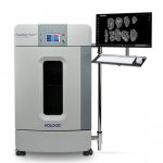
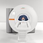
Siemens launches its smallest and most lightweight whole-body MRI
With its Magnetom Free.Max, Siemens Healthineers is presenting a new class of magnetic resonance imaging (MRI) systems that the company calls “High-V MRI.” The scanner’s unique combination of digital technologies and the new field strength of 0.55 tesla broadens the range of clinical applications for MRI systems. Magnetom Free.Max considerably improves pulmonary imaging with MRI and allows…