News • Deep learning & CNN

Algorithm differentiates small renal masses on multiphase CT

A deep learning method with a convolutional neural network (CNN) can support the evaluation of small solid renal masses in dynamic CT images with acceptable diagnostic performance.

Photo
67-year-old woman with 2.7-cm solid renal mass that was pathologically diagnosed as clear cell renal cell carcinoma after surgery. Unenhanced (A), corticomedullary phase (CMP) (B), nephrogenic phase (NP) (C), and excretory phase (EP) (D) CT images all show small solid renal mass correctly diagnosed as malignant lesion by CMP, triphasic (CMP, NP, and EP), and all-phase convolutional neural network models. Readers 1 and 2 also correctly determined small solid renal mass to be malignant (both 4 on 5-point scale).

The article was published in the March issue of the American Journal of Roentgenology (AJR).

Between 2012 and 2016, researchers at Japan’s Okayama University studied 1807 image sets from 168 pathologically diagnosed small (≤ 4 cm) solid renal masses with four CT phases—unenhanced, corticomedullary, nephrogenic, and excretory—in 159 patients. 

Masses were classified as malignant (n = 136) or benign (n = 32) using a 5-point scale, and this dataset was then randomly divided into five subsets. 

As lead AJR author Takashi Tanaka explained, “four were used for augmentation and supervised training (48,832 images), and one was used for testing (281 images).”
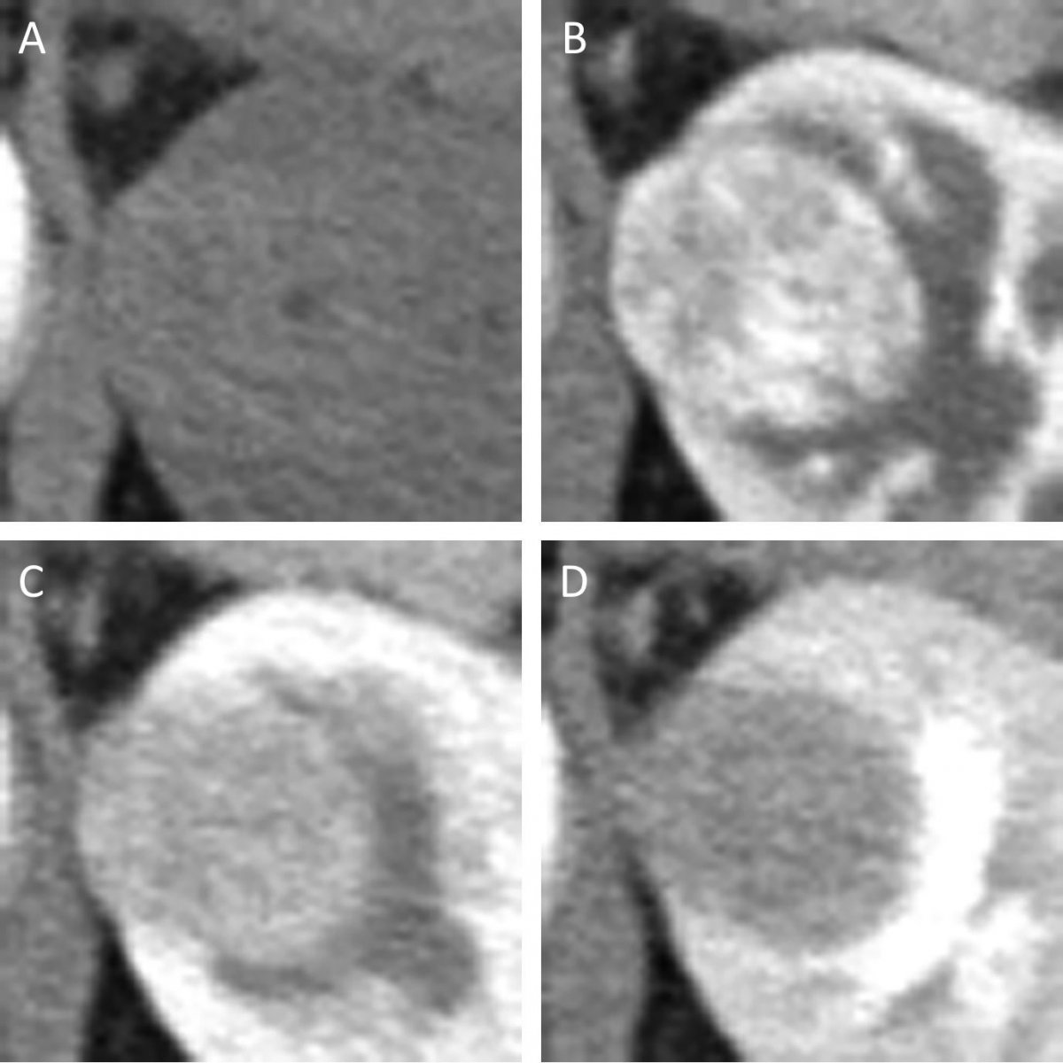
Photo
41-year-old woman with 2.6-cm solid renal mass that was pathologically diagnosed as angiomyolipoma after biopsy. A, Unenhanced (A), corticomedullary phase (CMP) (B), nephrogenic phase (C), and excretory phase (D) CT images show small solid renal mass correctly diagnosed as benign lesion by CMP convolutional neural network (CNN) model but not by other CNN models. Readers 1 and 2 incorrectly determined small solid renal mass to be malignant (4 and 5 on 5-point scale, respectively).

Utilizing the Inception-v3 architecture CNN model, the AUC for malignancy and accuracy at optimal cutoff values of output data were evaluated in six different CNN models. Finding no significant size difference between malignant and benign lesions, Tanaka’s team did find that the AUC value of the corticomedullary phase was higher than that of other phases (corticomedullary vs excretory, p = 0.022).

Additionally, the highest accuracy (88%) was achieved in the corticomedullary phase images. Multivariate analysis revealed that the CNN model of corticomedullary phase was a significant predictor for malignancy, “compared with other CNN models, age, sex, and lesion size,” Tanaka concluded.


Source: American Roentgen Ray Society (ARRS)

13.01.2020

Read all latest stories

Related articles

Photo

News • Algorithm-assisted diagnostics

AI in imaging: not as reliable as you'd think

Machine learning and AI are highly unstable in medical image reconstruction, and may lead to false positives and false negatives, a new study suggests. A team of researchers, led by the University of…

Photo

News • Imaging signatures

CT-based radiomics deep learning to predict lymph node metastasis in tumors

With a combination of radiomics and deep learning, researchers aim to noninvasively determine lymph node metastasis before surgery. This could lead to more accurate diagnosis and treatment strategies.

Photo

News • Smart diagnostic support

Brain imaging: bringing CT up to par with MRI

A new AI method for CT brain imaging may bring the modality to the level of detail usually reserved for MRI scans. This could enhance diagnostic support for conditions such as Alzheimer's disease.

Related products

Subscribe to Newsletter