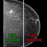
News • Screening performance
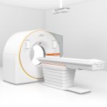
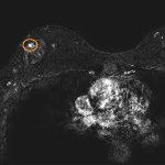
MRI reduces breast cancer deaths in women with 3 risk genes
Annual MRI screenings starting at ages 30-35 may reduce breast-cancer mortality by more than 50% among women who carry certain genetic changes in three genes, according to a new modeling analysis.