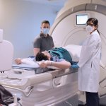
Medical display
JVC – CL-S1200
The CL-S1200 is a 30.9-inch color monitor with 12 megapixels for reporting medical images of various modalities such as CT, CR/DR, MR, ultrasound or mammography.


The CL-S1200 is a 30.9-inch color monitor with 12 megapixels for reporting medical images of various modalities such as CT, CR/DR, MR, ultrasound or mammography.
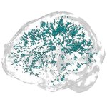
Researchers from Queensland have developed a new fMRI technique with vastly increased temporal resolutions, enabling them to capture the dynamics of brain activity at a sub-second level.
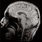
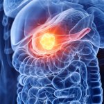
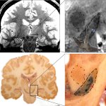
The distinction between primary tumors and metastases can be made quickly and accurately in brain tumors using radiomics and deep learning algorithms, a new study shows.
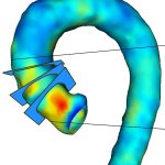
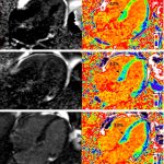
MRI-derived wall shear stress values predict pathological changes in the aortic wall in patients with ascending aortic dilatation. This can help identfy at-risk patients.

Medical imaging and healthcare IT company Esaote reports that it has completed the transition of its medical devices to comply with the new Medical Device Regulation (MDR).
For the first time, a new study has identified enlarged perivascular spaces in the brains of migraine sufferers. Results of the study will be presented at the annual RSNA meeting.
Using a special type of MRI, researchers have uncovered brain changes in patients up to six months after they recovered from Covid-19, according to a study being presented at the annual RSNA meeting.
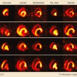
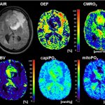
Swiss researchers developed a new MRI method to visualise metabolic processes in the body. Their objective is to improve the future diagnosis and treatment of heart disease.
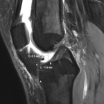
Magnetic resonance imaging (MRI) can reliably establish measurements for anterior cruciate ligament (ACL) “footprints” that are critical to the placement of grafts for reconstruction surgery.
Siemens Healthineers presented its latest magnetic resonance tomographs designed for clinical and scientific use at the company's Shape 23 Keynote: The 3T Magnetom Cima.X and 7T Magnetom Terra.X.
White matter hyperintensities (WMH) on the brain seen on MRI represent a biomarker associated with a 50/50 risk of death within five years after a first incident acute ischemic stroke (AIS) or transient ischemic attack (TIA).
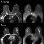
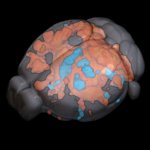
The risk of developing breast cancer is higher in breasts with high density. But why is that? Researchers at Linköping University have shown major biological differences that promote cancer growth.
To assess diffuse liver disease, MRI is currently the modality of choice. New developments in artificial intelligence (AI) could tip the scales in favour of CT imaging. At ECR 2022 in July, experts showed how AI technology enables CT to quantify liver fat as exquisitely as MRI.

Women with high-risk breast lesions (HRLs) and no family history of breast cancer or BRCA mutations are generally considered to be at moderate risk of developing breast cancer. Breast cancer screening guidelines suggest breast MRI be considered as a supplement to mammography. But is this expensive exam necessary?
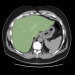
Pancreatic cancer tumours are being missed on CT and MRI scans, narrowing the window for life-saving curative surgery, research presented at UEG Week 2022 has revealed.
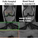
US scientists and engineers have found a way to improve the performance of traditional Magnetic Resonance Imaging (MRI) reconstruction techniques, allowing for faster MRIs to improve healthcare.
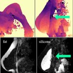
In MRI-based monitoring of silicone implants, separation of silicone and fat tissue is challenging. A newly developed algorithm was designed to assist in the task.
New method for the detection of alcohols combines zero- to ultralow-field nuclear magnetic resonance with the SABRE-Relay hyperpolarization technique.
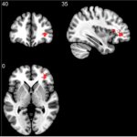
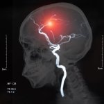
A new understanding of the 'swallow-tail', an anatomical landmark in the brain, could pave the way for earlier detection of Parkinson's disease.
The portfolio Philips presented at ECR 2022 revealed that the company not only advanced their products, but also listened to medical professionals and patients – and took their feedback to heart.
An advanced form of cardiac MRI has for the first-time enabled clinicians to measure the effectiveness of chemotherapy in patients with the life-limiting condition ‘stiff heart syndrome’.

Researchers are seeking alternatives to gadolinium-based contrast agents (GBCAs) to help raise levels of patient safety. An open hybrid session at ECR in Vienna heard how research across several centres has been examining the options of new approaches to reduce reliance on GBCAs.
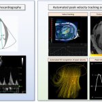
Researchers have developed cutting-edge imaging technology to help doctors better diagnose and monitor patients with heart failure. The state-of-the-art technology uses magnetic resonance imaging (MRI) to create detailed 4D flow images of the heart.
Using functional MRI with inhaled xenon gas, researchers have identified that long COVID symptoms are related to microscopic abnormalities that affect how oxygen is exchanged from the lungs to the red blood cells.
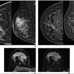
Contrast-enhanced mammography (CEM) may be a useful alternative test for neoadjuvant therapy (NAT) response assessment in patients with breast cancer who are unable to undergo MRI.