News • Radiophysiomics
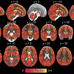
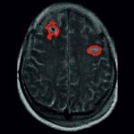
Diagnosing brain tumors using machine learning
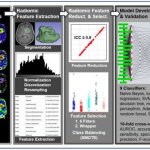
The classification of brain tumors—and thus the choice of optimal treatment options—can become more accurate and precise through the use of artificial intelligence in combination with physiological imaging.