Child diagnosed with abusive head trauma. He also had unexplained rib, humerus,...
Child diagnosed with abusive head trauma. He also had unexplained rib, humerus, and tibial fractures, three skull fractures, severe acute hypoxic brain injury with transtentorial herniation, and intracranial subdural hematoma in interhemispheric falx and tentorium. A. Sagittal STIR image from whole-spine MRI demonstrates edema in nuchal ligament consistent with ligamentous injury (arrow) and epidural hematoma extending along thoracic spine (arrowhead), associated with compression fractures from T4 to T7 (rectangle). Epidural hematoma isolated to thoracolumbar spine. B. Lateral radiograph of thoracic spine clinically interpreted as negative for spine fracture. C. Axial gradient-recalled echo T2-weighted MR image at L4 demonstrates posterior hematoma obliterating epidural fat, consistent with epidural hematoma (arrow).

Credit: American Roentgen Ray Society (ARRS), American Journal of Roentgenology (AJR)

News • Trauma imaging

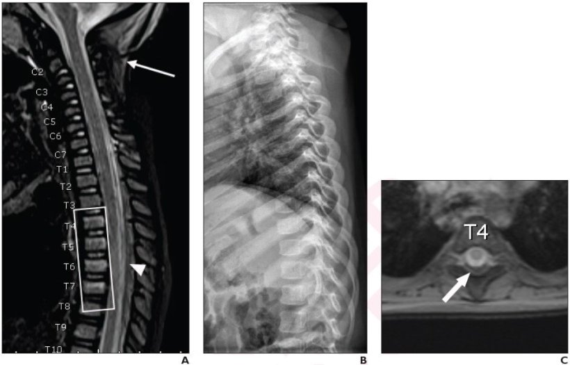
Suspected child abuse: Whole-spine MRI warranted

According to new research, whole-spine MRI commonly demonstrates isolated thoracolumbar injuries in children with suspected abusive head trauma.

"When performing spine MRI in children with suspected abusive head trauma, whole-spine MRI rather than cervical spine MRI may be warranted, to avoid missing isolated thoracolumbar injuries," clarified first author Boaz Karmazyn from Indiana University School of Medicine, Riley Hospital for Children.

Karmazyn and team's retrospective study included 256 children (170 boys, 86 girls; mean age, 5.9 months), who from January 2019 to December 2020, underwent skeletal survey and head MRI for suspected child abuse. Children with suspected abusive head trauma also underwent whole-spine MRI, per institutional protocol. Diagnoses for abusive head trauma were established via clinical record review, as well as injuries described in skeletal survey, head MRI, and head CT (if performed) reports.

Of the 148 total children with suspected abusive head trauma who underwent whole-spine MRI, 23.0% of examinations demonstrated injuries localized to the thoracolumbar spine. Injuries were localized to the thoracolumbar spine in 51.1% of examinations with major findings: Subdural hematoma, epidural hematoma, ligamentous injury, or fracture not identified by skeletal survey.

"This study represents the largest reported series to our knowledge of children with suspected abusive head trauma who were evaluated by whole-spine MRI," added the authors of the article in ARRS' American Journal of Roentgenology

Source: American Roentgen Ray Society

18.01.2022

Read all latest stories

Related articles

Photo

News • Pediatric cancer imaging

DW MRI measures tumor chemotherapy response with less radiation

Whole body diffusion-weighted magnetic resonance imaging (DW MRI) may aid in the assessment of cancer treatment response in children and youth at much lower levels of radiation than current…

Photo

Article • Photon-counting CT, strain imaging, 4D flow MRI

How new technologies shape the future of cardiovascular radiology

New approaches to cardiovascular radiology are evolving to help clinicians gain an increasingly better insight into heart conditions. Latest developments in cardiovascular radiology include…

Photo

News • Medical technology presentation

United Imaging showcases EU presence, AI-driven solutions at ECR 2024

United Imaging showcased its full portfolio of AI-empowered products at the ECR 2024 in Vienna. The company highlighted its significant EU growth since establishing its regional HQ in 2019.

Related products

Subscribe to Newsletter