News • New method
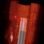
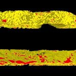
3D imaging to determine prostate cancer aggressiveness
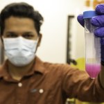
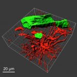
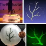
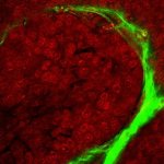
A team led by the University of Washington has developed a new, non-destructive method that images entire 3D biopsies instead of a slice. The 3D images provided more information than a 2D image — specifically, details about the tree-like structure of the glands throughout the tissue.