News • Diabetic damage
3D imaging shows how diabetes twists nerve fibers
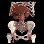
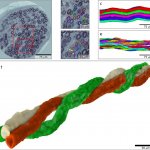
In an international collaboration led by Lund University in Sweden, researchers have used synchrotron light to study what happens to the nerves in diabetes. The technique shows the 3D-structure of nerve fibers in very high resolution. “This knowledge can be used to map mechanisms for how nerve fibers atrophy and grow back. It means that we can better understand how diabetes affects the nerves…