Image source: Shutterstock/IRINA SHI
News • Catheter ablation support
Software lets surgeon see real-time 3D map of heart
Innovative software that allows a surgeon to view inside a patient’s heart in real time while applying treatment has been used for the first time in the UK in operations at the National Institute for Health Research (NIHR) Leicester Biomedical Research Centre (BRC).
University of Leicester Professor and Leicester’s Hospitals Cardiologist Andre Ng used the new technique in operations on two patients suffering from atrial fibrillation (AF), the most common form of heart rhythm disturbance in the UK with more than 1.5 million people affected. The standard treatment for AF is catheter ablation, in which a long, thin tube is inserted into the heart via the femoral vein. The tip of the catheter creates scars (lesions) in the heart muscle, using either heat or cold, which disrupt the erratic electrical signals causing the AF. Applying the lesions at exactly the right points takes tremendous skill.
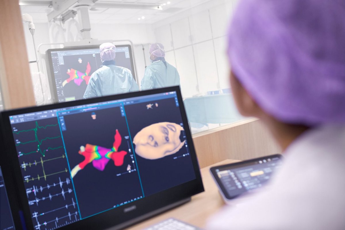
In August 2019, Professor Ng became the first cardiologist in the UK to use the KODEX-EPD cardiac mapping and navigation system in operations on two AF patients at Glenfield. The software uses electromagnetic technology to create real-time three-dimensional, high definition (3D-HD) images of the patient’s heart. Using the new system, Professor Ng was able to apply lesions with unprecedented accuracy and precision, thereby increasing the operation’s effectiveness and reducing the risk of complications. Both operations were successful and the patients have now been discharged.
“Catheter ablation is an established treatment for AF but the results are variable,” said Professor Ng. “An Achilles heel of this therapy is that we do not have real-time information of how good the ablation lesions we deliver are, which is largely regarded as the Holy Grail of the procedure. The KODEX EPD mapping system uses a novel di-electric energy source which allows us to electrically image unique characteristics of the heart and other structures, like a high fidelity radar system, thereby providing unique advantages over other mapping systems currently used. We are very pleased to be able to have access to this new technology at an early stage and hope to consolidate this through further research work to explore and develop its potentials, which we hope will be translated to improved procedure outcomes.”
The KODEX EPD software is part of a new multicentre study, the Patient Specific Outcome Trial (PSOT). Glenfield Hospital is the UK’s lead for this multicentre international trial.
Source: University Hospitals of Leicester NHS Trust
02.09.2019