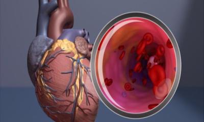
Image source: MRC-LMS; from: Schweitzer R, De Marvao A, Shah M et al., Radiology: Cardiothoracic Imaging 2025
News • exCMR study
What biking in an MRI can tell us about heart health
New research from the MRC Laboratory of Medical Sciences (LMS) will help more doctors use a simple test called Exercise Cardiac MRI to diagnose heart conditions accurately and at an earlier stage.
A quarter of deaths in the UK are caused by heart disease, the equivalent of one person every three seconds. Improving diagnostics will allow for earlier diagnosis and better health outcomes.
Research published in Radiology: Cardiothoracic imaging and led by the computational cardiac imaging group at the LMS has established the normal range of measurements (reference ranges) that indicate someone’s heart is responding in a healthy way to exercise. These ranges are needed to better interpret a medical imaging technique called Exercise Cardiac MRI (exCMR), which means this work could help increase uptake of the test in clinics across the UK and internationally.
If we make them exercise during their MRI they can no longer compensate and we get a much clearer idea of who has the least resilience and who is really at risk
Declan O’Regan
The team carried out live cardiac imaging in 161 healthy people between the ages of 22 and 77 during exercise to determine what the response of a healthy heart looks like. Importantly, the team found how a healthy response to exercise differed between men and women. Men’s hearts were more responsive to exercise than women’s, even allowing for body size, which needs to be accounted for when diagnosing heart conditions.
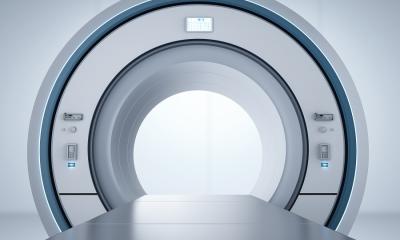
ExCMR is used to study the heart’s response to exercise and is a valuable tool for diagnosing and managing various cardiovascular conditions. A special attachment allows you to pedal like you’re on a bike while you lie in the MRI scanner. As you pedal the team can observe your heart respond, allowing them to get a clearer idea of your heart health.
For some people, an early or undiagnosed heart condition doesn’t cause many problems while they’re resting. The signs of their condition become more apparent as they carry out their daily activities such as walking up the stairs or running for the bus. Usually, people undergo an MRI scan while they are in a resting position. “Young people and people in the early heart stages of heart disease are very good at compensating for their condition when they’re at rest,” says Professor Declan O’Regan, head of the computational cardiac imaging group at the LMS, “But if we make them exercise during their MRI they can no longer compensate and we get a much clearer idea of who has the least resilience and who is really at risk.” This means that by imaging the heart during exercise we could diagnose heart disease earlier and more accurately. Better and earlier diagnosis will lead to better outcomes for patients with heart disease.
Recommended article

Article • Focus on radiology
Magnetic resonance imaging (MRI)
Imaging without ionising radiation: MRI uses magnetic fields to look inside the body. Keep up-to-date with the latest research news, medical applications, and background information on MR imaging.
While the exCMR is known to be a useful test, it’s not widely used in clinical practice. “One of the barriers to more clinics using this test is that until now we didn’t know what normal and abnormal responses to exercise looked like,” says Declan, “Our study provides reference ranges for normal health so we can now interpret the results from this test and get a better idea of where people sit on this range.”
The test is relatively inexpensive and is safe and straightforward to use. Now, having a better understanding of reference ranges will help more doctors to use it. “This study will help clinicians better understand the value of this test and when to use it. This is an important step in getting the exCMR into practice,” says Declan.
Operating at the interface between fundamental research and clinical translation, this work closely aligns with the LMS’ strategy to work towards understanding the mechanisms that underlie human health and disease which could lead to new or improved diagnostics and treatments.
Source: Medical Research Council (MRC) Laboratory of Medical Sciences; by Emily Armstrong
08.06.2025