Image source: Catharina Ziekenhuis
News • Vascular disease assessment
New ultrasound technique to predict aneurysm rupture
Researcher Floor Fasen, who is based at e/MTIC – a collaboration between the Catharina Hospital, Eindhoven University of Technology and Philips, is working on a smart way to better predict whether a weak spot in the abdominal artery (aorta) could be dangerous.
In the future, her technique could help to treat patients earlier and in a more targeted way with less burden.
An aneurysm is a dilation in a blood vessel, usually in the aorta, the largest blood vessel in the body. Often it does not lead to any symptoms, but if the blood vessel ruptures, it is life-threatening. Such a weak spot is now often discovered by accident during a scan for something else. If an aneurysm is detected, this is followed up by additional check-ups to see if the dilation increases. "Doctors now mainly look at the growth and diameter of the aorta," says researcher Floor Fasen. "However, sometimes a small aneurysm ruptures, while a large one does not. So, perhaps there are better ways to assess risks."
Many people with an aneurysm feel like they are walking around with a ticking time bomb. Especially if you know it can tear, but don't know when or if it will ever happen
Floor Fasen
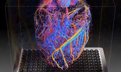
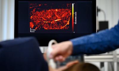
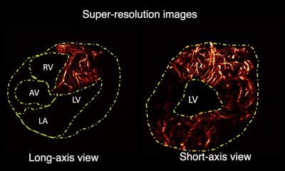
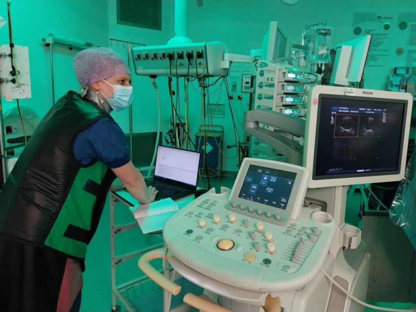
Fasen has developed a method with which she can look at the wall of the aorta from the inside. She achieved this during operations in which a stent was moved through a small opening in the groin to the weakened spot in the aorta. Once in place, the stent is deployed and serves to decrease the pressure of the flowing blood on the vessel in that region, so that the fragile vessel wall is spared. During this so-called Endovascular Aneurysm Repair (EVAR) procedure, she temporarily inserted a thin tube into several patients. At the end of the tube is a small ultrasound probe, which is like a pregnancy ultrasound. "This allowed me to measure the wall thickness of the aorta," Fasen says. "This doesn't work though with an ultrasound through the top of the abdomen, as my colleague Esther Maas did as part of her work. However, my method was more invasive, which makes it only suitable for a small group of patients: people who were already undergoing EVAR surgery."
The measurements carried out by Fasen yielded a considerable amount of data. Fasen is now using this data to analyze and train an AI model that automatically recognizes features in the aortic wall. "Right now, this model measures the wall thickness. Then we try to make connections between wall thickness, calcifications, and blood clots," explains the PhD researcher. "Calcifications are hard segments in the otherwise flexible vessel, and they can restrict the natural movement of the wall. We think this can increase the risk of cracks in the wall as a result."
To train the AI model, Fasen first practiced the implantation process in the lab using pig aortas, which are very similar to human aortas. "I built a sort of fake belly of gelatin, in which I placed the pork aortas," she says. "This creates a realistic environment for the ultrasound to operate and approximates how it is in the human body."
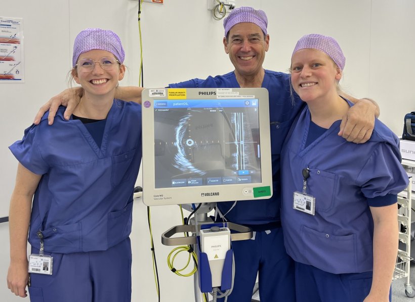
Image source: Catharina Ziekenhuis
Although the AI model cannot yet predict for a specific patient whether an aneurysm will rupture, patterns are visible, and Fasen and her colleagues are learning a lot about the growth of aneurysms and changes in the vascular wall. "In the future, of course, you hope to be able to say something for each patient individually, but that is still difficult to do with this invasive method. Yet, what we have already learned is valuable. The next researcher working on this project will be able to build on the insights we now have into how wall thickness, calcifications, and clots are related to aneurysm growth."
Two scientific papers have now been published about Fasen’s research: one on simulating ultrasound images for training purposes and a second paper on measurements taken from pig aortas. A third publication - based on the measurements in patients - is currently being reviewed by experts. Fasen is in the final phase of her PhD research. "The writing of my dissertation is in full swing. I hope to finish by the end of this year and then have my PhD defense in the spring of 2026."
The hope is that in the future doctors will be able to better predict whether someone is really at risk of an aneurysm. This would help avoid any unnecessary scans and hospital visits, as well as relieve stress and tension for patients. "Many people with an aneurysm feel like they are walking around with a ticking time bomb," says Fasen. "Especially if you know it can tear, but don't know when or if it will ever happen."
An even more advanced development is the idea of a 'smart plaster': a kind of ultrasound sticker that you can stick on your stomach and continuously take images of part of the body. "Our research group is experimenting with this right now," says Fasen. "However, it will only be really useful in practice in a few decades. Companies such as Philips could be the ones to develop it commercially."
Source: Eindhoven University of Technology (adapted from Catharina Hospital)
19.08.2025