

Image source: Siemens Healthineers
Sponsored • How to deliver more and better care at lower costs
Moving the needle in MRI productivity
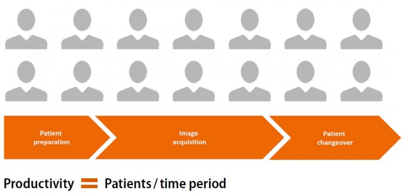
In an industry where every second and every click counts, workflow inefficiencies consume as much as a third of the MRI procedure time.1 This is a key area of focus where technology advances can radically change what is possible with an MRI exam. Given declining reimbursements, fewer skilled resources, and the system-wide burden of chronic diseases, maximizing productivity is a strategic goal for a healthcare organization to reach its optimal performance. The best way to achieve this goal is the holistic optimization of the entire imaging value chain: From patient preparation to image acquisition and postprocessing – while maintaining a high standard of image quality as well as patient comfort.
Less than one minute for a more reproducible and automated patient preparation2
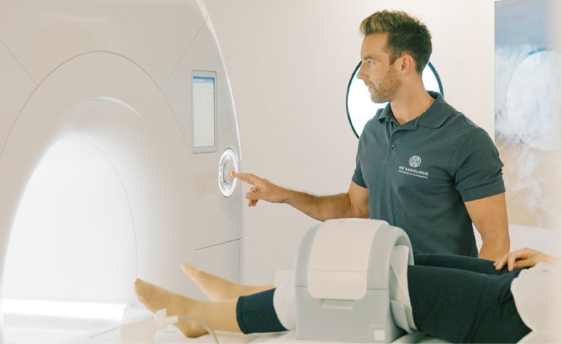
Patient preparation for MRI is a complex task: Technologists need to manage software and hardware. It is during the patient-facing part of the process where a positive patient experience is crucial: One study found that 42 percent of surveyed adults stated they were afraid of undergoing an MRI.3 The more comfortable the patient and the better their experience during patient set-up the easier it is to get the scan right the first time.
Innovations such as BioMatrix technology improve consistency, enhance patient comfort to avoid motion and accommodate challenging physiology and anatomy. Integrated Respiratory Sensors, for example, detect breathing patterns from the moment the patient lies on the table to help the system anticipate motion and secure a high-quality image, all while reducing patient set-up time.
BioMatrix Select&GO leverages artificial intelligence to help the technologists position patients faster and avoid repositioning delays –
no matter how tall, big, or mobile a patient is. The BioMatrix Dockable Tables with eDrive allow the technologist to move a patient quickly and easily to and from the scanner; one patient can be set up while another is in the scanner, thus improving workflow.
Up to 50 percent time savings in image acquisition4
Image acquisition is a critical phase in multiple aspects of the diagnostic workflow, including tailoring the MRI exam to specific patients, improving the patient experience, and image quality. Exam consistency across multiple scans and follow-ups for the same patient supports a more accurate read.
myExam Companion guides the acquisition or even automates it to large degrees. This improves productivity, standardization, and consistency across patients and in follow-up exams, independent of the skill level of the technologist. This results in 45 percent5 lower tasks at hand for the MRI technologists versus using a non-myExam Companion Workflow. Acceleration technologies such as Simultaneous Multi-Slice (SMS) and Compressed Sensing (CS) make the acquisition faster than ever. When patients cannot hold their breath, acceleration technologies can expand access to specific MRI applications for some of the sickest patients.
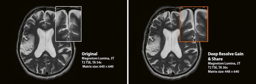
New advances, such as Deep Resolve6, are planned to provide an advanced reconstruction technology using deep learning and Artificial Intelligence. Its components Deep Resolve Gain and Sharp are designed to make scans faster and boost workflow efficiency while improving patient comfort.
Faster post processing due to AI-powered features
While the number of MRI exams is continually on the rise, the workload of each radiologist is increasing dramatically. On average, a radiologist interprets one image every 3–4 seconds, 8 hours a day.7 Software innovations, including Artificial Intelligence, allow for faster post processing and eliminate errors and inconsistencies.
AI-Rad Companion is an AI-powered radiology assistant that supports by performing automatic measurements and preparing
the results in the form of valuable clinical images and quantifications for MR Brain and Prostate.
Improvements in clinical practice
Die Radiologie, a large multi-site radiology practice in Munich, Germany, is already using the Siemens Healthineers MRI productivity solutions, performing a broad range of MRI examinations. Magnetom Sola has enabled Die Radiologie to reduce the average slot time to 20 minutes, resulting in a throughput of 30 patients in a 10-hour shift. Thanks to this accelerated workflow, Die Radiologie has increased patient throughput by 17 percent while maintaining excellent image quality.
References:
1 Beker K, et al. AJR Am. J. Roentgenol. 2017; 209: 836-844
2 Data on file
3 Siemens Healthineers survey conducted in May 2015 in which 2,000 UK adults were asked about their attitudes on their health, hospitals, and medical appointments
4 Data on file
5 Case Study by Prof. Forsting, Prof. Antoch, Department of Diagnostic and Interventional Radiology and Neuroradiology, University Hospital, Essen, Germany
6 Deep Resolve is still under development and not commercially available yet. Its future availability cannot be ensured
8 The statements by Siemens Healthineers' customers described herein are based on results that were achieved in the customer’s unique setting. Since there is no “typical” hospital and many variables exist (e.g., hospital size, case mix, level of IT adoption), there can be no guarantee that other customers will achieve the same results. This statement is from a person whose institu-tion is engaged in a collaboration with Siemens Healthineers
9 Data on file
15.04.2021


