Image source: Shutterstock/Levent Konuk
News • Vital imaging
Study: contrast MRI after a heart attack could increase survival
A new study from the University of Surrey and University College London has revealed that treatment for heart attacks could be improved thanks to a novel method of evaluating heart function using contrast-based MRI scans.
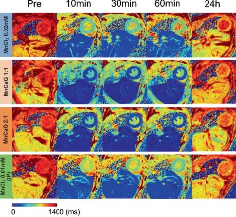
According to the British Heart Foundation, heart and circulatory diseases cause more than a quarter (27 per cent) of all deaths in the UK, which equates to more than 160,000 deaths each year – or one death every three minutes. The research, published in the journal Advanced Science, found that injection of the trace mineral manganese could enhance MRI scans so that they provide more accurate details of heart function than traditional MRI methods.

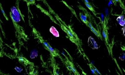
Image source: Jasmin et al., Advanced Science 2021 (CC BY 4.0)
These findings, if confirmed in human subjects, could have major implications for the treatment of heart attack patients. The findings could also be of great use in the preclinical evaluation of treatments for patients who suffer from cardiac ischemia – a reduction in blood supply to the heart muscle that could lead to cardiac arrest.
The study also suggests that if manganese-enhanced MRI is performed within the first few hours of a heart attack it could be used to determine the optimal treatment regime for individual patients – helping to regulate changes in the cardiac muscle and thereby further improving survival chances. Findings were evaluated by examining the infarct size and blood supply at three key intervals: one hour, one day and 14 days after a myocardial infarction was induced.
Dr Patrizia Camelliti, Principal Investigator and Senior Lecturer in Cardiovascular Science at the University of Surrey, said: "Magnetic resonance imaging (MRI) is increasingly used to diagnose and give information on heart conditions. This research using mice allows us to measure the health status of the heart muscle rapidly after a heart attack and could provide important information for optimising treatments in patients”.
Source: University of Surrey
12.05.2021


