Article • Expanding potential
Low-field MRI: Less is more
Currently, two opposing trends can be observed in MRI: on the one hand 1.5T scanners are increasingly replaced by 3T scanners for standard clinical MRI applications. On the other hand scanners with lower and even significantly lower field strength have become commercially available in the past two years. A session at this year’s European Congress of Radiology (ECR 2021) took a closer look at low-field MRI scanners with about 0.5T and ultra-low-field MRI scanners with a field strength of 0.05T.
Report: Michael Krassnitzer
‘The key challenge of those low and ultra-low-field MRI systems is the poorer signal-to-noise ratio,’ said Professor Andrew G Webb, Director of the Centre for High-Field MRI at the University Hospital Leiden (Netherlands). The signal-to-noise ratio (SNR) of a 1.5T scanner is six times higher than in a 0.55T system and even 300 times higher than in a 0.055T system. Each MRI system aims to deliver a good ratio of true signal (i.e. reflecting actual anatomy) to unavoidable background noise. While low-field and ultra-low-field MRI scanners struggle with SNR, they do offer certain advantages over standard MRI scanners.

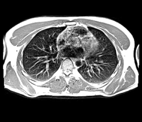
Image source: Siemens Healthineers/University Hospital Erlangen
Webb pointed out the areas where low-field MRI scanners receive top scores: firstly, the low purchasing and site costs. Secondly, low-field MRI is less susceptible to artefacts caused by metal implants and unwanted effects caused by air-filled cavities (lungs, nose). The specific absorption rate (SAR) is about one tenth lower than in 1.5 T scanners, the wave lengths of the magnetic waves is above the critical value for implants. ‘Therefore, we don’t need to be worried about counter-indications,’ Webb underlined. Since gradient acoustic noise is weaker, noise stress for the patient is significantly lower. Moreover, low-field MRI offers advantages with regard to relaxation times: T1 relaxation time is significantly shorter than in 1.5T scanners while T2* relaxation time is significantly longer.
With its Magnetom Free.Max, Siemens Healthineers set a new standard. The 0.55T system, launched last year, is suited to examine lung, abdomen, brain and cardiovascular structures. In addition, image-guided interventions can be performed. ‘This is basically a standard MRI scanner,’ explained Professor Matthieu Sarracanie from the Department of Biological Engineering at the University of Basle. The system’s benefits are light weight (3,200 kg) and a compact design with the large bore and closed helium-free cooling system. The drawbacks are the poorer resolution compared to 1.5T systems and that the system requires the same space and infrastructure as a standard scanner.
Image source: Siemens Healthineers
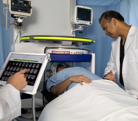
In the ultra-low-field MRI segment the Hyperfine scanner, launched in 2019, aims to be the new benchmark with a field strength of 0.054T. Weighing just 630 kg and being the size of a nightstand this mobile system can be used at the bedside, primarily in emergency settings. No site costs, no specific power supply, so specific cooling, even fewer artefacts caused by medical implants, no undesirable side effects caused by air – all are advantages, according to Webb.

Image source: Hyperfine Research Inc.
‘The magnetic waves are so long that the critical length for implants is 75 metres, which is irrelevant for clinical purposes,’ the physicist pointed out. ‘This system has enormous potential,’ Sarracanie, a co-founder of Hyperfine. He also pointed at the low costs of approx. $30,000 plus a monthly fee. A dedicated site is no longer needed; the carbon footprint is comparatively small and the system can be powered by a standard wall outlet. However, he also points out drawbacks: the system is tailored for specific exams. Currently only brain and extremities can be scanned. Moreover, the Hyperfine is a proprietary system which means, for example, that images can only be stored in the company’s own cloud.
Open-source projects make MRI available for everyone
However, there are also non-commercial ultra-low-field systems. With his University Hospital Leiden team, Webb built a 0.05T portable system which is used for neuroimaging. ‘Rather than using several large and heavy magnets we use many small ones – each about the size of a one-cent coin,’ he explained. The dice-shaped 12 mm magnets are mounted on acrylic glass rings – 23 altogether with 2848 magnets. Costs: €4,000 plus a few thousand euros more for gradient and RF coils and amplifiers. ‘This is an open-source project. Anybody who is interested can build such a scanner based on our construction plan.’ These systems are also designed for one specific exam, for example paediatric hydrocephalus.
The system was designed primarily for use in developing countries. ‘Seventy percent of the world population don’t have access to MRI,’ Webb pointed out. ‘Due to low costs, mobility and standard power supply ultra-low-field MRI scanners can now make MRI available all over the globe.’
ESR/EFOMP – High-field vs low-field MRI: time for a re-think?, ECR 2021, Vienna 4/3/2021
20.05.2021


