Article • Imaging with benefits
Low-field MRI: good quality images, few artefacts and high patient comfort
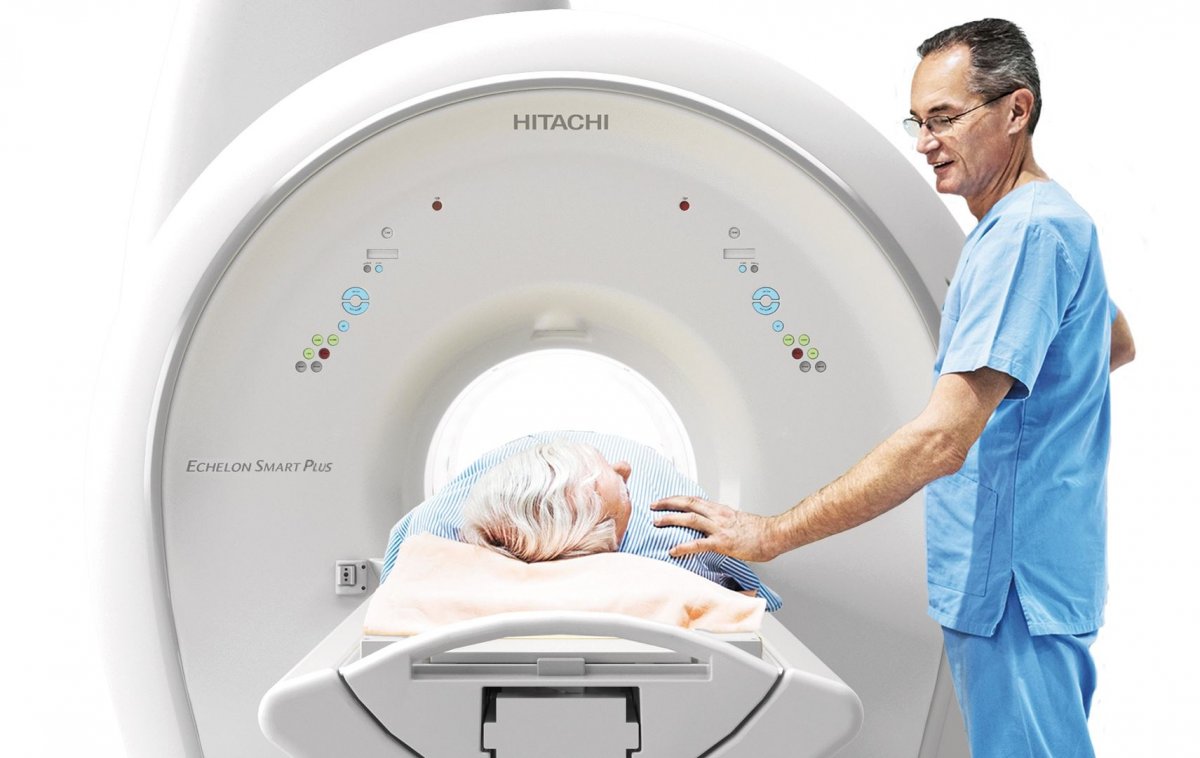
A crucial advantage of low-field MRI is the fact that these open systems are suitable for claustrophobic patients – after all one out of twenty patients are concerned.
Report: Michael Krassnitzer
Low-field MRI is the flavour of the season after several 1.5T models were launched. The major advantages of this technology are a relatively low purchasing price and operating costs and few artefacts while the image quality is comparable to a 1.5T scanner. Patient comfort is another benefit of low-field scanners. This advantage, however, tends to get short shrift despite the fact that it might indeed be the crucial benefits from the patient’s point of view. An industry session at the European Radiology Congress (ECR 2021) dealt with the issues surrounding low-field MRI. Geared towards the user, neither developers nor scientists were on the panel but owners of private MRI practices.
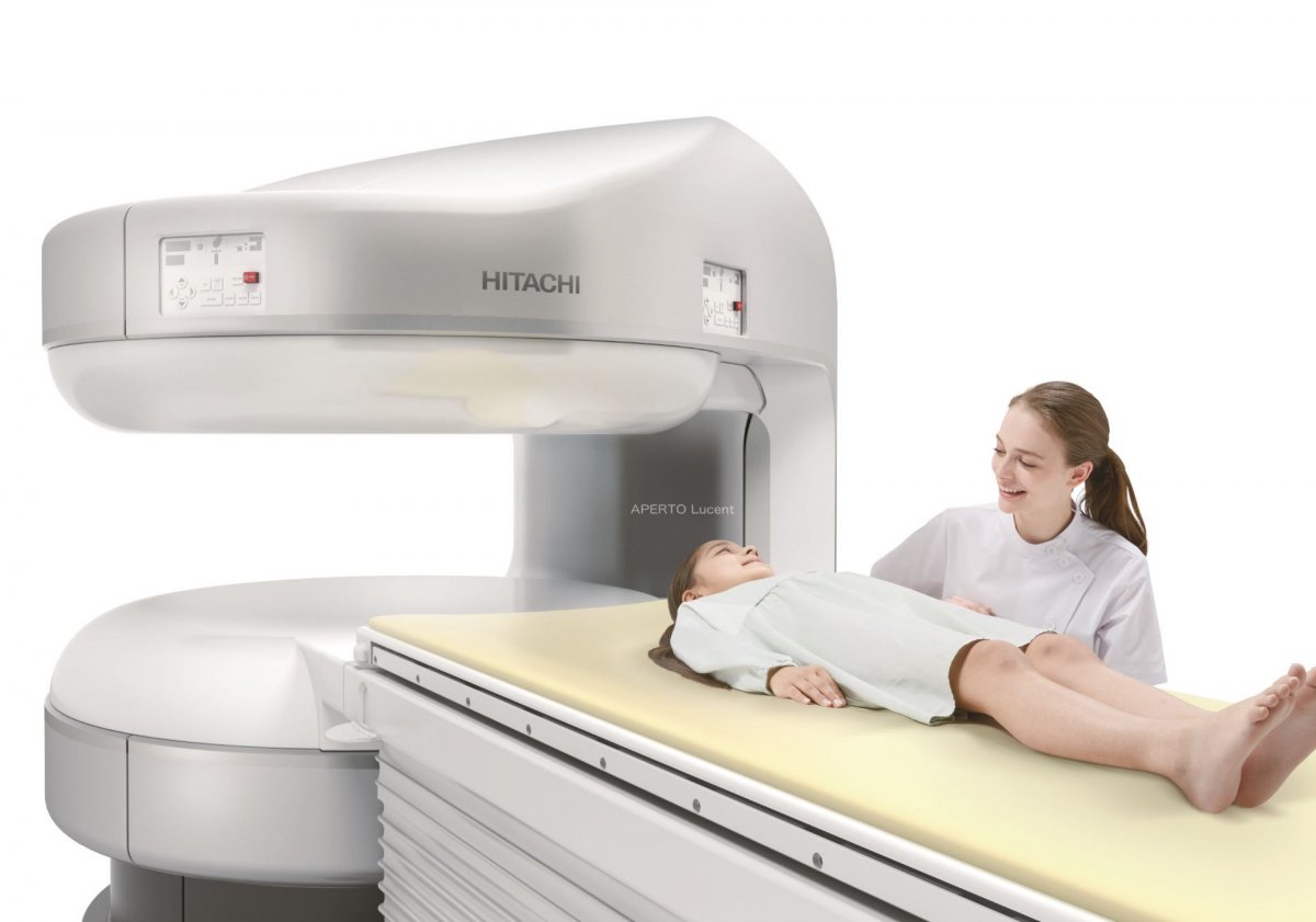
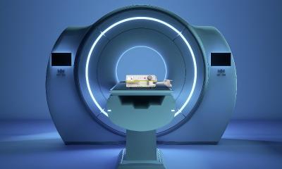
A low-field scanner is an open MRI system where the patient is not in a closed tube. For obese and claustrophobic patients this is the only option to get an MRI exam. According to the four radiologists on the session panel, roughly five percent of all MRI patients suffer from anxiety and panic attacks in small or enclosed spaces. These persons are unable to stand the narrow tube of a conventional scanner and require full anaesthesia. “In some patients claustrophobia is so severe that they can’t even enter the changing room,” reports Dr Jan Veryser, who is operating an MRI centre in Sluis (Netherlands). For the physicians these five percent – that is 1 out of 20 – claustrophobic patients are a major issue.
“A lot of attention, love and time,” is Veryser‘s approach to the claustrophobic patients. “When the patients make their appointment we ask them to have a look at our scanners to understand that there are no tubes. Before the exam, even before they enter the changing rooms, we show them the scanners again so they can see it.” The radiologist is present during the entire exam to provide support. Moreover, the patients can take along a person they trust and who holds their hand during the scan. Even the design of the room takes the needs of claustrophobic patients into consideration: there are no small changing cubicles but one large room and the distance from the reception area to the exam room is a mere 10 m.
Dr Carsten Figge, head of a radiology practice in Paderborn (Germany), focuses on claustrophobic patients. His strategy includes the pleasant design of the scanner, sophisticated lighting and ceiling windows that open up to the sky. Moreover, the staff is well trained to support patients before, during and after the exam. “We aim to make the stay as pleasant as possible for our patients – from the first to the last second,” Figge underlined. This is more important to him than the technical specifications of the low-field scanner: “The number crunching is irrelevant.” Nevertheless, he did compare his open 1.2T with his 1.5T and reported that the 1.5T scanner yields slightly better results than the 1.2T scanner for prostate exams. In musculoskeletal imaging and neuro-imaging however he did not notice relevant differences. In breast imaging the open scanner offers the advantage that women can lie flat on their belly.
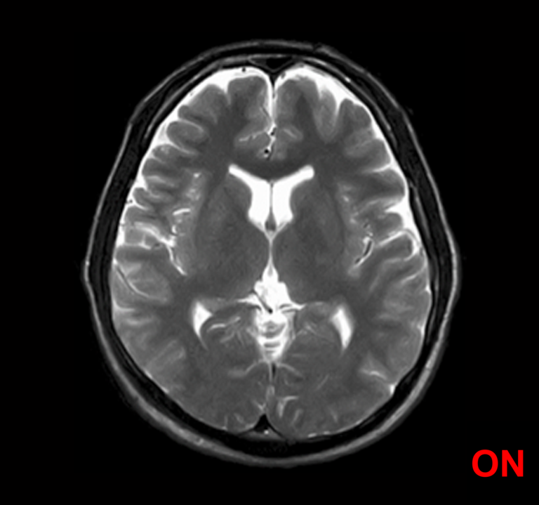
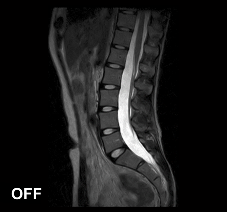
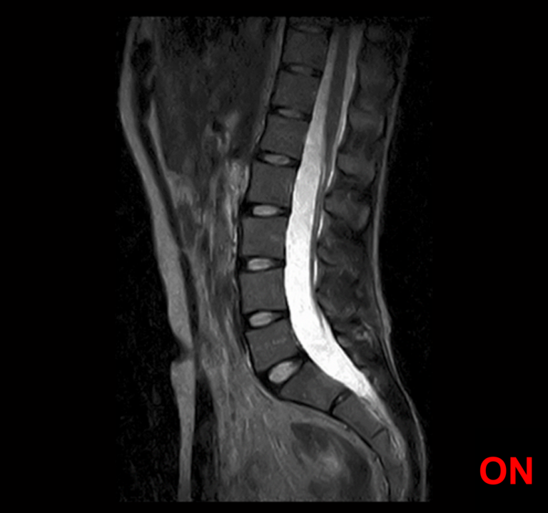
Dr Mihaly Aradi, director of a private radiology practice in Pécs (Hungary), uses 0.4T as well as 3T systems. “With low-field MRI you need to schedule more time for an exam,” he said and added that “the new digital signal processing technologies compensate weaker signals well.” In his experience, 0.4T scanners are as good for routine exams as the 3T system. “For MRI scans of the spine both systems deliver pretty much identical quality,” the Hungarian radiologist said and suggested that indeed “every clinic that specializes in spine surgery should have a 0.4T scanner as well as a 3T scanner.” He also pointed out that the low-field scanner’s advantage is the reduced number of artefacts: “Distortions are so minute that structures are clearly visible even when they are next to metal implants.”
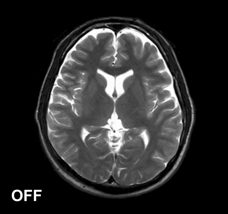
Image comparison: MRI scans with Lucent Plus' IP-RAPID functionality ON/OFF
And last but not the least, Dr Liliana Rechmeier, who co-heads a radiology practice with her husband in Bad Neuenahr (Germany), stressed the low operating costs of a low-field MRI scanner. As owner of a private practice, she said, she has to keep the costs under control: “Due to the low costs for servicing and electricity and since no liquid helium cooling is required the operating costs are only about 60 percent of those for a conventional scanner. Over the course of eight to ten years of use, that’s a substantial amount of money.” And in view of the good image quality she urges her colleagues to rethink current prejudices: “Low-field MRI is no amateur scanning; it delivers really good radiological images.”
The battle of fields in MRI, ECR 2021
*with the AI powered SynergyDrive Automation Suite
20.05.2021