Source: Norfolk and Norwich University Hospital (NNUH)
News • Imaging
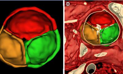
4D flow MRI scans to help patients with heart disease
A cutting-edge imaging technique that creates 4D flow images of the heart has been carried out for the first time in the UK at the Norfolk and Norwich University Hospital (NNUH).
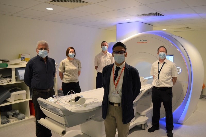
Cardiology patients at NNUH have become the first to benefit from ongoing research that uses magnetic resonance imaging (MRI) to create detailed images to diagnose heart valve disease. Dr Pankaj Garg, NNUH Honorary Consultant Cardiologist and UEA Norwich Medical School lecturer, is putting his research into clinical practice for the first time, thanks to the latest MRI equipment installed as part of an £8m project to replace ageing CT and MRI scanners at the Trust and new 4D flow software.
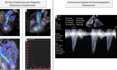
Using the latest state-of-the-art MRI at the Trust and new 4D flow software, a six to eight minute non-invasive scan and an hour of processing provides precise imaging of the heart valves helping doctors determine the best course of treatment for patients.
Dr Garg, who has been working on imaging technology innovations to benefit heart disease patients since 2014, said that the standard method of diagnosing heart valve disease is with ultrasound method called echocardiography, which is limited to only one-directional flow imaging in the heart. “It is very exciting to be able to do 4D flow scans for our patients, which is more precise and if we know more precisely where the leak is in a heart value, we are able to make better informed decisions. Every patient who has their scan this way is having one of the most cutting-edge scans in the country and the patients are quite excited to be getting this expertise.”
Dr Garg hopes to train more clinicians in this novel clinical technology and work with leading industry partners to speed up the time it takes to process the huge amount of data to create the detailed imaging.
Heart and circulatory disease, also known as cardiovascular disease (CVD), causes a quarter of all deaths in the UK, and is the single biggest condition where lives can be saved by the NHS over the next 10 years. Dr Garg is the first at NNUH to receive a Wellcome Trust Clinical Career Development Fellowship Award with a funding of £746,198 to support ongoing international collaborative research for the development of non-invasive tools to predict pressures in the heart.
Source: Norfolk and Norwich University Hospital (NNUH).
19.06.2021