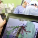
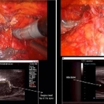
MRI

Photo

MRI: Still the best tool for seizures

Anti-seizure medication is not successful in all patients, while in others such medication can have side effects. In recent years significant technical advances have delivered better imaging results, which, combined with growing demand for a surgical solution from patients whose medications do not control seizures, or those not wanting to take medication constantly, has led to an increase in…

Photo

Europe’s first AIRIS Vento LT

November saw the opening of the private Avicenna Clinic in Berlin, a clinical "gem" set in the heart of Berlin, not far from the famous Kurfürstendamm. This spinal hospital is also the first in Europe to have installed the AIRIS Vento LT, Hitachi Medical Systems’ latest generation of open MRI systems in its AIRIS Series.

Photo

New driver of innovation and efficiency in radiology

Philips is ushering in a new era in radiology science with Imaging 2.0, a concept fueled by integration of technology, clinician and patient. Showcasing its commitment to pioneering innovative, cost-effective solutions, Philips is highlighting technologies that focus specifically on the patient, in addition to advanced networking tools that facilitate greater collaboration between radiologists…

Photo

Ingenia, the first-ever digital broadband MRI system

At the 96th annual meeting of the Radiological Society of North America (RSNA) in Chicago, Philips showcased Philips Ingenia, the first-ever digital broadband magnetic resonance imaging (MRI) solution. Driven by Philips’ commitment to developing innovative patient care solutions, Ingenia MR delivers exceptional image clarity, scanning efficiency and scalability. The Philips Ingenia MR system is…

Photo

MPR 3-D echocardiography

A cardiologist at a UK hospital has become the first in the world to develop a technique to ‘slice’ 3-D images of the heart into intricate sections using computer software. The method, devised by consultant congenital cardiologist Dr Joseph Vettukattil at Southampton General Hospital, is known as multiplane review (MPR) 3-D echocardiography. This allows cardiologists to identify heart defects…

Photo

The Virtual Autopsy Table

This is imaging with a ‘wow’-effect: The Swedish Centre for Medical Image Science & Visualisation (CMIV) in cooperation with the Norrkoping Visualisation Centre has developed a ‘Virtual Autopsy Table’ that allows a unique look inside the human body and takes interaction with volumetric medical data to a new level.

Photo

Modern radiological diagnostics for the evaluation of muscle diseases

Muscular diseases belong to a heterogeneous group with various causes like neurogenic, metabolic, dystrophic, or inflammatory mechanisms as well as channelopathies leading to disorders of the muscle cell membrane potential. In most progressive disease cases the result is a focal or general muscle weakness that, unfortunately, is a very unspecific symptom. Standard neuromuscular literature…

Photo

Diffusion weighted whole body MRI

Malignant diseases rank second in mortality rates in Germany. These patients thus receive a major proportion of ambulant and hospital care, with apparent socioeconomic consequences. To optimise treatment planning, for all solid tumour entities it is mandatory to delineate or stage the primary extent of tumour invasion and spread prior to therapy as precisely as possible.

Photo

Article • MRI

9-, 10- or even 11-Tesla – is more always better?

Advancing magnetic resonance imaging (MRI) up the Tesla scale may sound good, but will it produce the results and patient safety radiologists actually desire? Faced with the question: ‘How many Tesla should it be?’, Professor Siegfried Trattnig MD, head of the Centre of Excellence in high-field MRI at the University Clinic for Radiodiagnostics, Medical University of Vienna, and Austria’s…

Photo

The potentiated value of MR-PET imaging

Even more precise diagnoses, even better process controls -- the future of MR-PET technology has dawned. The first commercial, full-body hybrid scanners are either waiting in the wings or already installed. But what does the introduction of the MR-PET really mean for clinical practice? Professor Heinz-Peter Schlemmer MD, Head of the Radiology Department at the German Cancer Research Centre in…

Photo

Stroke - New options for prevention and therapy

It’s hardly surprising that stroke was a focus at the 20th meeting of the European Neurological Society (ENS) -- every year at least a million people suffer one in the EU. In an interview with Bettina Döbereiner, Professor Karl Max Einhäupl, Director of the Department of Neurology at the Charité - University of Medicine Berlin, of which he is Chairman of the Board and co-chair of the ENS…

Photo

MRIs may lead to unnecessary breast surgery

Women could be undergoing unnecessary breast surgery as a result of having magnetic resonance imaging, says an expert on bmj.com. In the last decade, says Malcolm Kell, Consultant Surgeon and Senior Lecturer at the Eccles Breast Screening Unit at University College Dublin, MRI or magnetic resonance mammography has become the most favoured type of investigation for high risk patients when combined…

Photo

Investigating sports injuries

Radiologist Dr Karl-Friedrich Schünemann works in a group* practice in the small German city of Paderborn. Focusing on sports injuries and using MRI, CT and X-ray, the specialist medical team provides services such as neuroradiology, orthopaedics and CT-guided pain therapy. ‘The fact that we count a number of professional athletes among our clients also attracts many amateur athletes who…

Photo

Aortic valve replacement in the third dimension

No white lab coats anywhere; instead men in hard hats, equipped with hammers and drills. The Düsseldorf University Hospital’s Cardiology Pneumology and Angiology Clinic is a construction site, but once the workmen have packed up their tools and removed the scaffolding the view to the human heart will be unobstructed and clearer than ever before. Here, innovative patient care and a highly…

Photo

Multi-modality imaging in coronary artery disease

The focus on Coronary artery disease – from genes to outcomes at ESC 2010 underlines the importance of CAD diagnosis on the scientific agenda. During the session ‘Multi modality imaging to detect coronary artery disease’, chaired by Professor JJ Bax, of the Cardiology Department at Leiden University Medical Centre, the Netherlands, experts will discuss, for example, the advantages of the…

Photo

Getting to the heart of things

Not only is heart failure one of the single biggest causes of morbidity and mortality in man, but the incidence of the condition is steadily increasing. Rising to this challenge, innovative medical diagnostic techniques with ever greater performance are constantly being introduced so that early, unambiguous detection of the underlying condition is now possible, enabling the prompt initiation of…

Photo

The ultra-high-field MRI symposium

Early problems of ultra-high field Magnetic Resonance Imaging (MRI) have been overcome by successful development of adequate hardware. In consequence big efforts have been achieved in structural imaging, as well in functional imaging. Basic scientists and physicians who work in ultra-high-field MRI in Europe and the USA, met at the Berlin Ultra-high-field Facility (BUFF), in the Max Dehlbrück…

Photo

Instead of the “Tube” – An Innovative MRI is open for all Patients

Modern magnetic resonance (MR) tomographs with very strong magnetic fields produce images of steadily increasing precision and are currently gaining more and more importance in radiological diagnostics. In conventional MRI the patient lies in a tunnel, which many people find difficult or even impossible to do, given a scan time of approximately 20 minutes. Physicians of the Joint Radiology…

Photo

1st in UK - the 1.2 Tesla open vertical high-field MRI system

During the opening of the fully renovated InHealth MRI Centre at Mayday Hospital in Croydon, UK, three renowned Harlequin rugby players demonstrated just how ‘open’ the newly installed Oasis 1.2 Tesla highfield open MRI system is. Rugby players are notably large. This Hitachi system can accommodate patients who cannot fit into a conventional closed bore type of magnet.

Photo

Open for all patients

Hitachi Medical Systems UK has been awarded the contract for OASIS™ 1.2 Tesla high-field open MRI by InHealth MRI Centre at Mayday Hospital, Croydon – the high performance system with a truly open architecture available in the UK. This unique product combines unrivalled patient comfort together with first class imaging comparable to that of a closed bore system.

262 show more articles
Subscribe to Newsletter