Iterative reconstruction
A field report from the user’s viewpoint
About two years ago iterative image reconstruction was officially introduced for CT imaging. Since then, no other technological innovation has raised more hope that the dose of X-ray based, cross sectional imaging can be significantly lowered. The possibilities of this procedure have not yet been exhausted.
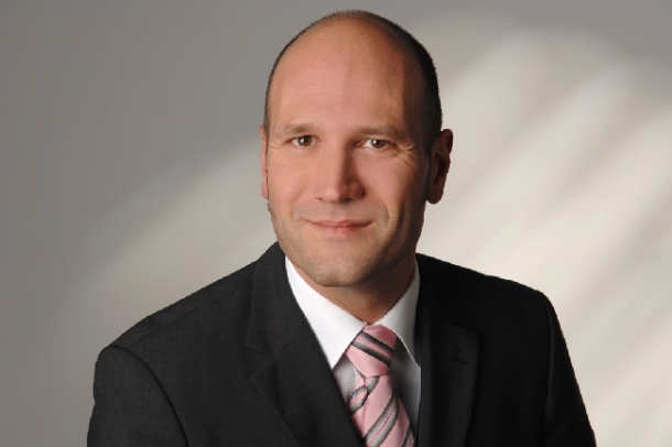
What then is the current state of play in iterative reconstruction, and where are developments in this field heading? Seeking and update, Karoline Laarmann spoke with Dr Stefan Wirth, computer scientist and radiologist at the Institute for Clinical Radiology at Munich University Hospital.
Asked what iterative reconstruction does, Dr Stefan Wirth explained that CT images are basically overlays of real information and image noise. ‘This image noise consists of an unavoidable part, such as tissue homogeneities and quantum mottle, as well as an avoidable part of noise which results from less than exact calculations. This new innovation specifically tackles this last component by minimising its ratio and therefore increasing image quality, or, more importantly, facilitating a radiation dose reduction.’
Why have the mathematical calculations not been exact, so far?
SW: The FBP (filtered back projection) algorithm, which has been used for last 30 – 40 years for the retrospective calculation of the image data from the value of attenuation, is based on a large number of very simplified assumptions. It is, for example, based on the assumption that the radiation source and the detector are both punctiform. In reality, these components are actually of a defined size. On the other hand, the FBP algorithm assumes a right-angle beam incidence. With the detector arrays these days being many centimetres wide, this is also not geometrically correct.
How does the new procedure solve this problem?
The initial approach is iterative construction (IR), which uses a noise pattern and calculates image data step by step, separates image information in one part and noise in another and then minimises the latter based on the pattern until the point where it falls below the level of tolerance.
The manufacturers offer different varieties here. IR is possible with a comparatively small additional amount of computing time and lasts a maximum of two minutes longer than FBP. The different manufacturers’ products differ mainly in that some of them (IRIS-Siemens, AIDR-Toshiba) work exclusively with the data in the image space, whilst others (ASIR-GE, IDOSE-Philips, SAFIRE-Siemens) additionally work with the raw-data space. Whilst the first group works like a filter-based smoothing, an optimisation step in the second group additionally also effects a change in the raw data.
Independent of that, the norm is to present an overlay of start and end data in the final image set. Based on existing experience, IR makes it possible to save between 30-60% of the required dose. Mostly manufacturers also offer a retrospective upgrade for existing CT scanners with this technology, which presents a good alternative and encouragement for users to benefit from it as early as possible.
What other approaches are being followed?
I’m convinced that the additional consideration of exact geometry with the so-called model-based iterative reconstruction (MBIR) will shape the future of CT to such an extent that it is likely, at one stage, to be perceived as such a great advance in technology as the introduction of spiral CT or modulated MD-CT.
However, at the moment there is only one manufacturer who offers this technologically completely new procedure, and that is GE Healthcare with their VEO product. However, taking into account the actual characteristics, such as X-ray tube focus and beam/detector geometry, makes MBIR very intensive regarding the computing time required. Even with extremely powerful, and therefore expensive, servers currently this image reconstruction still takes half an hour. Moreover, MBIR cannot retrospectively be installed on older machines.
So, do you still see this solution as extremely promising?
Definitely. We have tested the product for half a year and will include it in our clinical routine from this year. In studies we have already seen that the image quality improvement from FBP to IR is around a quarter of the image quality improvement from IR to MBIR.
Additionally, MBIR processes the data set as a complete volume, which, based on existing experience, has very positive effects on artefacts in particularly problematic regions of the body. Different image convolution kernels are no longer necessary for MBIR. The really big advance in technology is therefore to be expected with MBIR. However, image reconstruction must still become much faster so that it can become widely used in clinical practice.
14.02.2012