UK researchers are working on a new MRI technique
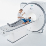
UK researchers are working on a new MRI technique called hyperpolarised MRI – or Dynamic Nuclear Polarisation (DNP) – that can utilise more of the available nuclei than traditional MRI, helping to overcome some of its limitations by increasing sensitivity 10,000-fold or more. DNP is part of a longer-term aim to improve cancer mortality with the help of novel cancer imaging tools.