The end of blooming effects
Anatomic and functional cardiac examination with CT scanning
During the RSNA 2011, Professor Uwe J Schoepf MD, was asked what will be the chosen procedure of the future in cardiac imaging, he answered without hesitation: ‘Definitely CT,’ and, the Director of Cardiovascular Imaging at the Medical University Charleston, South Carolina.
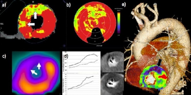
perfusion examination (a, b, d, and e) shows lack of perfusion (arrows) in the anterior
wall of the cardiac muscle in good correlation with the results of the nuclear-medical
SPECT examination (c). The flow of blood in healthy and diseased tissue can be measured and compared with good precision (d). The examination also faciliates the assessment of the patiency of the bypass (e).
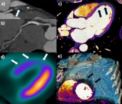
interventricular artery (a) as well as the open stent in the right coronary artery (b). The newly occurring occlusion causes reversible ischemia of the anterior wall of the heart (arrows in c, d, e), which could be detected with the stress dual energy CT in a comparable manner to the SPECT (d), but in the context of the overall thoracic anatomy (e).
He added: ‘We are trying to transfer as many examinations as possible that are currently carried out with MRI or NUM, to CT.’ The professor is convinced that CT will soon meet all the requirements of a ‘one stop shop’ and it will be possible to answer structural, anatomic as well as functional questions with the help of just this one modality.
MRI diagnostics is considered the international gold standard for the functional imaging of cardiac perfusion – ‘although,’ he says, ‘this procedure is not actually the most common examination used for this purpose in any particular country, worldwide.’ In the US, nuclear-medical procedures such as SPECT are far more likely to be used for this purpose. Beyond these procedures, Prof. Schoepf is convinced that it will be CT examination that will become established as the only non-invasive procedure for the examination of coronary arteries. The one stop shop CT modality will cover all aspects of coronary disease. Coronary narrowing or occlusion is already routinely diagnosed via CT, but the same modality also makes it possible to determine myocardial blood flow.
Dual energy CT is the preferred procedure in Prof. Schoepf’s clinic, to assess coronary arteries or the patency of bypasses as well as of the heart muscle, particularly through the direct determination of the myocardial blood volume. The merely indirect determination of the myocardial perfusion via Nuclear Medicine (NUM), the resulting false positive findings and the insufficient categorisation may soon be outdated.
However, the necessity of two examinations to detect reversible ischemia – a stress-related lack of myocardial perfusion – remains, but these can be performed using only one modality! After the first rest CT, a second image is taken under pharmacologically induced hyperaemia. Although the associated higher cardiac frequency pushes the CT to its limits when examining the coronary arteries, a contrast medium injection at the maximum stress point facilitates a good assessment of the heart muscle under stress in the second CT.
Therefore, the MRI examination remains Prof. Schoepf’s preferred procedure only for the examination of myocardial perfusion. However, as soon as an examination of the coronary arteries is called for, the CT can be used for both - and without the interpretation of images based on their intensity scales as carried out with the MRI. This becomes particularly obvious with the dual energy CT, which offers many opportunities of image reconstruction. With this procedure one basically looks directly at the iodine in the heart muscle via material differentiation, and the real composition of the cardiac tissue becomes apparent.
CT is also superior to MRI when it comes to the quantification of blood flow: Whilst there is no exact, linear connection between important, and not only in his institute. There are worldwide efforts to increase this and other potentials of CT – in cardiological as well as radiological working groups. For Prof. Schoepf this also means that CT cannot be considered the speciality of either cardiology or radiology, but that those with the most experience of this type of examination must always carry it out. In his clinic in Charleston it happens to be the radiology department, at Johns Hopkins it is the cardiology department.
Worldwide, the majority of CT scanners are operated by radiologists, which, going forward, should ensure a strong involvement of radiology in the most modern procedures of cardiac imaging. Unfortunately, not everyone shares Prof. Schoepf’s enthusiasm for CT as the method of choice for the examination of coronary disease, certainly not medical insurers. In the US, and in most centres in Europe, it is still mainly considered a research field. In Asia, however, CT is being fully reimbursed by insurers in the same way as NUM used to be – which it has now completely replaced. For Prof. Schopf, this is further ‘ammunition’ for ‘his’ CT.
21.02.2012