News • Tissue tension measurement
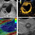
New ultrasound method could lead to easier disease diagnosis
A new ultrasound method that can measure the level of tension in human tissue for the first time - a key indicator of disease - has been developed by researchers from the University of Sheffield.