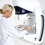
Imaging

Photo

When B Mode has an Image Problem

Ever heard this from a patient? 'Wouldn't it be great if it was in colour.' It's true; B mode lacks the glamour of Doppler. We're guilty of it ourselves, we love Doppler. How frequently do we hear 'did you put colour on that'? When was the last time you asked 'how much temporal smoothing did you use'? And yet every time I watch others scan I witness Sonographers scanning from various angles,…

Photo

Dynamic Contrast Enhanced Ultrasonography (DCE-US)

Early functional evaluation of new treatments in oncology is of major importance. Overall survival rate is the best criterion for assessing treatment, but unfortunately it calls for lengthy follow-up whereas treatment efficacy must be ascertained as soon as possible. The morphological criteria normally used (WHO or RECIST) do not lend themselves easily to new therapies which often induce lesion…

Photo

Smooth workflow, excellent diagnostics and patient-friendly handling

With 715 beds, the University Hospital Maastricht is one of the leading hospitals in the Netherlands. The modern building offers 22 operating theatres and in 2005 24,362 patients were admitted. Due to rotating staff systems and multidisciplinary responsibilities in diagnostic imaging departments in large hospitals like the University Hospital Maastricht, the workflow has to be as easy as possible…

Photo

Carotid Plaque Imaging

Ischaemic stroke accounts for 80-85% of all cerebrovascular accidents and causes considerable morbidity and mortality, thereby placing a significant burden on western societies. In the UK alone, stroke costs amount to 10.4 billion EUR annually. These events commonly are a consequence of systemic atherosclerotic disease and with the internal carotid arteries supplying 75% of cerebral blood flow,…

Photo

Endoscopic Ultrasound Transducer with FNA Biopsy Capability

Endoscopic Ultrasound with Fine Needle Aspiration Biopsy (FNAB) capability is relatively new for Toshiba. Recently the company has introduced a transducer enabling gastroenterologists to perform these kinds of examinations. Dr. Paul Fockens (PhD), Director of Endoscopy at the Academic Medical Centre (AMC) of the University of Amsterdam, is an expert in this field. He has been Director of…

Photo

Clinical Experience with a Superconducting MRI Scanner

Following the installation of a 0.5-T superconducting magnetic resonance imaging (MRI) system at our hospital in 1987, we have been performing a large number of examinations as a key regional hospital serving the city of Muroran, Hokkaido, and surrounding areas. However, compared with more recently developed MRI systems, 0.5-T systems tend to have poorer image quality and a restricted range of…

Photo

X-Rayed Masterpieces

Even today, 105 years later, the X-rays have not lost their fascination and importance. The discovery of X-rays in November 1895 by the physicist Wilhelm Conrad Roentgen introduced the 'X-ray era' and caused great change in all areas of life. One of the most fascinating fields of research is the analysis of works of art by means of X-rays. The 'anatomical structure', i.e. the composition of the…

Photo

Technical Insights into the Dual-slice CT

In 1999, the Aquillion/MULTI and Asteion/MULTI multi-slice systems with highly integrated 34-channel detector system, both consisting of a total of 30,464 detector elements, were introduced. On the basis of such a structured detector matrix, a new image reconstruction procedure was used. This so called MUSCOT (Multislice Conebeam Tomography) procedure made the dream of an isotropic voxel in the…

Photo

Procedure and Clinical Value of CT Fluoroscopy

The past several years, interventions using ultrasound for real-time image monitoring in diagnostic and therapeutic procedures have increasingly been integrated into the daily clinical routine. Limitations due to image-distorting physical effects, such as the total reflection at the soft tissue/lung boundary, are associated with the use of ultrasound. CT fluoroscopy on the other hand enables…

Photo

Tissue Doppler Imaging for assessment of pseudonormalization of the mitral inflow pattern

Impairment of left ventricular relaxation may lead to pulmonary congestion and symptoms of heart failure even in patients with preserved systolic function. Doppler echocardiography has become the non-invasive method of choice for the assessment of both systolic and diastolic dysfunction. However, Doppler-derived parameters such as mitral inflow velocity, deceleration time and isovolumic…

Photo

Contrast Harmonic Imaging (CHI)

Case 1 50-year-old male with a colorectal cancer. Greyscale imaging clearly shows the lesion, as an echogenic 3 cm mass (Fig.l a), which must be considered as suspicious for a metastasis from his colorectal cancer but benefit of the doubt must be given. Colour Doppler (Fig. 1b) is not helpful. At 33 seconds after SonoVue injection (early sinusoidal phase), Vascular Recognition Imaging (VRI)…

Photo

CT Fluoroscopy

In recent years, interventional procedures using realtime image control by means of Ultrasound have been increasingly integrated into the clinical routine as diagnostic and therapeutic procedures. However, a series of limitations in ultrasound result tram physical effects that interfere with the image. Combining fluoroscopy with a computed tomograph (CT) results in a procedure that enables…

Photo

Application of Vascular Recognition Imaging (VRI)

Vascular Recognition Imaging (VRI) is a low MI broadband colour Doppler method that images the interactions of Sonovue with microbubbles in a non-destructive manner as perfusion images of a quality never before achieved. The perfusion image represents the low velocities of unmoving or barely moving microbubbles as green-coded tissue perfusion, while the higher velocities of the veins and arteries…

Photo

The Perfect Partnership

The Ultimax with image intensifier & CCD camera technology is already well established as a premium multipurpose digital imaging end interventional system. The addition of DynaDirect FPD technology with its exceptional high-resolution performance simply makes the Ultimax the most advanced high performance multipurpose digital imaging system available. The combination of the Ultimax and…

Photo

Contrast Harmonic Imaging at its best

Over the last years Toshiba has actively participated in the development of Contrast Harmonic Imaging. Around 1996 one of the first agents that became available on the European market was Levovist TM (Schering AG, Berlin, Germany). This was a galactose-based contrast agent. Levovist could be used as a Doppler signal enhancer throughout the entire vascular system. With the development of these…

Photo

Aquilion 16 - A new CT concept in optimized patient care

Two university hospitals in Germany and Japan were the first worldwide to receive the Aquilion 16 high resolution computed tomograph (CT) from Toshiba Medical Systems for simultaneous recording of 16 body slices with extremely high image resolution. A computed tomography Aquilion 16 was put into operation on October 7, 2002 at the central campus of Charite Berlin (Figure 1). This is the third…

Photo

Dose Efficiency Index (DEI)

Image quality has been the most important criterion in CT scanner development for both radiologists and manufacturers, due to the diagnostic value of these systems. Recently, considerable concern has been expressed about the radiation dose to patients from CT examinations because: - CT delivers a higher dose than conventional film-screen examinations - Technological advances in CT have…

Photo

From head to toe

Without doubt, the B-mode imaging is the most important examination method in prenatal ultrasound. Well over 90% of detectable congenital anomalies can be recognised using the two-dimensional realtime imaging method. Hence, paramount importance is certainly ascribed to the quality of the B-mode image in an ultrasound system. In addition, other ultrasound modalities help us to considerably enhance…

Photo

Interventional Neuroradiology

In Johns Hopkins therapeutic radiology, we perform 2,600 procedures in neurointervention and 14,000 peripheral interventions each year, totalling over 16,000 image-guided procedures a year in 14 rooms. We changed our approach to image guided therapy in June 2001, when we installed a new O.5 mm multidector CT (Aquilion, Toshiba America Medical Systems) and two 3D visualization workstations (Vital…

Photo

Looking at a New Generation

Most babies are born healthy, but there is still a small risk for all women to give birth to a baby with some type of anomaly. Ultrasound has proven to be one of the most reliable procedures to prenatally diagnose some of these conditions. Ultrasound can be used during the entire pregnancy, but in the first trimester ultrasound examinations are, as a rule, used to exactly calculate the pregnancy,…

Photo

New Image Guided Therapy Suites at Sick Kids

In May 2001, the world's first CT-XR hybrid suite for image guided therapy in paediatrics was put into operation. The new Centre for Image Guided Therapy (IGT) at The Hospital for Sick Children allows surgeons and interventional radiologists to do something that's never been done before - work side-by-side when diagnosing and treating children. A total of 4 interventional suites were equipped…

Photo

The Perfect Partnership

The Ultimax with image intensifier and CCD camera technology is already well established as a premium multipurpose digital imaging and interventional system. The additional DynaDirect FPD technology with its exceptional high-resolution performance simply makes the Ultimax the most advanced high performance multipurpose digital imaging system available. The combination of the Ultimax and…

Photo

Optimization of Temporal and Spatial Resolution for Cardiac CT Diagnostics

Further developments in computer and detection technology over the last few months and years have made it possible to overcome, to a large degree, the previously limitations of medical computer tomography in spatial and temporal resolution, and thus to permit “detail-true” representation of the beating heart. The diagnostic workflow and computer-tomographic diagnosis of the heart using the…

Photo

Advancing Pediatric Imaging with 16-Slice CT

Pediatric imaging presents many unique and difficult challenges to the radiologist. Correct and rapid diagnosis is extremely important in pediatric imaging and to that end we constantly strive for tests that offer more information, accuracy and versatility. However, it is also our duty to provide tests that are safe and minimise exposure to ionizing radiation. In recent months there has been…

Photo

Aquilion 32

With the launch of the Aquilion 32, Toshiba Medical Systems Corporation continues at the forefront of computed tomography (CT) technology. The most advanced 32-slice CT scanner available, the Aquilion provides increased patient comfort and the most flexible choice of speed and accuracy in CT scanning.

116 show more articles
Subscribe to Newsletter