From head to toe
Philips high performance CT opens up new possibilities in diagnostics
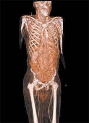
The new Philips 256-slice Brilliance iCT came in to use recently at the University Hospital in Ulm. The system produces quick, high-res scans with 80% less radiation.

The pin-sharp images promise new possibilities for cardiac diagnosis and treatment, as well as research into severe cardiac disease. ‘We now have state-of-the-art technology that will open up new opportunities in the clinical field as well as in research,’ said Professor Hans-Jürgen Brambs.
‘The 256-slice will be primarily a cardiac scanner for us,’ explained Dr Martin Hofmann, head of the hospital’s radiology department. ‘Speed and resolution are of equal importance for the quality of examination results. These two components will enable us to achieve fast, reliable diagnoses.’
As the heart moves continuously, an examination scan used to be carried out during the ‘quiet’ phase, based on ECG readings, which prevented motion artefacts on the images. However, cardiac image acquisition is now so fast that good clinical results at the lowest possible dose can be achieved even at high cardiac rates. In the future, the accurate display of unprecedented details of the coronary vessels will be possible — and it will even be possible to recognise the circulatory phases
of the myocardial tissue.
With the Brilliance iCT scanner radiation exposure for the patient is up to 80% lower than with systems used today. This is possible, among other reasons, due to the high speed of the equipment, so the patient is exposed for much shorter periods of time, and because of the highly sensitive detector, which is better able to record image data and can process them at lower doses.
To meet the increasingly complex clinical specifications, Philips engineers completely redesigned the CT platform. This included a novel type of X-ray tube, a specially developed detector and unique, pneumatic suspension technology for the imaging components that rotate around the patient. ‘The specifically developed suspension technology can be compared with that used in hovercrafts,’ explained Dr Gerald Poetzsch, who heads the CT business sector at Philips. ‘This is the only technology that allows the gantry, which weighs several tons, to rotate around the patient four times per second. The resulting centrifugal forces, which affect the individual components, are enormous. At this high speed of rotation, one ton of weight turns into 35 tons – comparable with the weight of a large truck.’
Thanks to the combination of these new developments it is now possible to achieve detailed and clear images of an entire organ within seconds, including those of the heart and brain, and long-term mutations can also be detected at a very early stage. Apart from cardiac examinations, the iCT in Ulm will also be used for clinical research on other organs. ‘The combination of morphological, angiographic, dynamic and functional imaging opens up new possibilities,’ said Dr Hoffman. ‘We hope that the system will facilitate significant progress in the fast diagnosis of strokes and in the highly sensitive detection of internal bleeding in seriously injured accident victims.’
30.04.2008
- angiography (117)
- CT (605)
- display (438)
- imaging (1641)
- markets (545)
- medical technology (1555)
- tissue (226)


