Cardiac software launched for CS Thin Client
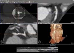
Visage Imaging has launched its Cardiac Analysis software for the Visage CS Thin Client, which offers 'advanced visualisation and quantitative analysis for cardiac CT studies, such as calcium scoring, coronary artery analysis and left ventricle analysis,' the firm reports.
‘The Visage CS Thin Client/Server is a scalable advanced visualisation solution that provides fast and consistent access to all patient images, anywhere inside and outside the hospital, across multiple interconnected sites, and through the internet.’
The cardiac software tools are fully integrated into the workflow and technology platform so that
it can be accessed on all client computers – a dedicated cardiac workstation is not necessary. Visage CS Cardiac Analysis allows viewing and post-processing of even the largest multi-phase cardiac CT studies at the click of a mouse, Visage reports.
‘Visage Thin Client solutions, like the cardiac one, enhance the entire clinical workflow, with advanced tools for 2-D, 3-D and 4-D image review and interpretation, post-processing, data management and image distribution. The original CT and cardiac CT data from multiple potential sources can be managed and archived consistently in a single system across the entire hospital enterprise. That is why we are talking about a cost-effective visualisation tool that optimises workflow not only within one department but over the boundaries e.g. of radiology and cardiology,’ explained Colin Murphy, Vice President of Sales & Marketing at Visage Imaging.
The new software provides, for example for left ventricle analysis, automatic short/long axis reformatting, segmentation of left ventricle with a few clicks, a global and by segment presented ejection fraction and wall motion analysis by segment. And, Visage adds, ‘For coronary artery analysis new features include automatic tracing with only a few clicks, a powerful vessel visualisation, an easy to use cross-sectional measurement and stenosis calculation, as well as a curved reformatting for efficient assessment of vessel lumen and adjacent structures.’
30.04.2008