Sponsored • Tomosynthesis
Launching a new field of investigation
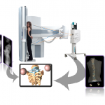
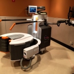
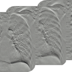
Tomosynthesis is an advanced application that allows a multi-slice acquisition and provides a reconstruction of a volume. Several acquisitions at low dose are acquired with a single sweep of the X-Ray tube around the region of interest.