News • Neuroimaging
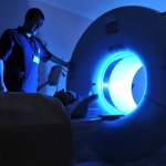
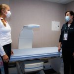
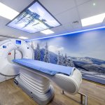
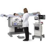
Stroke detecting: Portable MRIs almost as effective as stationary MRIs
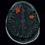
Portable MRI machines detected ischemic strokes, or strokes caused by clotting, in 90% of patients scanned.


Portable MRI machines detected ischemic strokes, or strokes caused by clotting, in 90% of patients scanned.
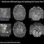
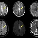
Cedars-Sinai physician-scientists are pioneering imaging techniques and investigating new biomarkers to improve multiple sclerosis (MS) diagnosis and treatment.
Patients with neurological conditions such as dementia or autism can prove especially challenging for radiographers. A session at the ECR Overture in March gave insights to a patient-focused approach.
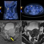
Diagnostic imaging in women’s health advances: PET/CT might provide a more accurate imaging alternative to CT in ovarian cancer. T2*-weighted MR imaging in deep endometriosis detection also shows promise, but ultimately falls flat.
Researchers from Charité – Universitätsmedizin Berlin have shown that massive electrochemical waves in the brain act as a marker announcing an impending ischemic stroke.
An algorithm built to assess scar patterns in patient heart tissue can predict potentially life-threatening arrhythmias more accurately than doctors can.
In the second year of the pandemic, JVCKenwood also draws a positive conclusion despite all difficulties. 'We were also able to welcome some highlights in 2021, both on the part of new monitors and new customers,' says Marcel Herrmann, Marketing Manager Medical Imaging. 'Nevertheless, the situation remains tense here and there.'
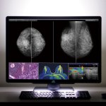
A major new study shows that artificial intelligence (AI) is a promising tool for breast cancer detection in screening mammography programs.

Contrast agents in the wastewater and power-hungry imaging systems: The eco-footprint of healthcare is huge, and radiology departments are among the main culprits. An expert panel at the ECR Overture explored ways to make the field “greener”.
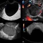
The appearance of ovarian lesions on ultrasound is an effective predictor of cancer risk that can help women avoid unnecessary surgery.
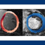
Synthetic correlated diffusion makes cancerous tissue glow in medical images could help doctors more accurately detect and track the progression of cancer over time.
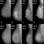
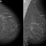
A software based on deep learning with convolutional neural networks can accurately and consistently classify breast density on mammograms according to BI-RADS criteria.
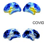
A significant number of Covid-19 neurological complications – such as fatigue, headache, and cognitive impairment – are ultimately reversible, according to new research.
Scientists at the Spanish Center for Cardiovascular Research (CNIC) have led the development of a new 3D ultrasound method that improves the assessment of cardiovascular risk in healthy individuals.
Hologic and the Women's Tennis Association (WTA) kicked off a multi-year partnership, launching a joint vision to achieve greater wellness and equality for women.

A mathematical analysis of data obtained with a MRI approach can identify brain cell damage in people at early stages of Alzheimer's.
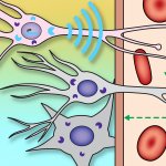
Using a novel probe for functional magnetic resonance imaging, researchers have devised a way to monitor individual populations of neurons and reveal how they interact with each other.

When it comes to imaging stable cardiac chest pain, which modality should be used as the first-line test to investigate coronary artery disease: CT or MRI? Radiologists discussed the strengths and limitations of the two approaches in a lively Pros and Cons session at ECR Overture.
To bridge new equipment installations in radiology, temporary high workloads, or long-term additional capacity, Canon Medical provides mobile solutions according to customers’ needs and wishes.
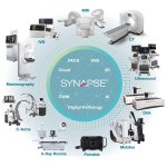
Fujifilm aims to become an entity that can contribute to the advancement of human health by supporting the ongoing transformation in the healthcare industry – as a “One-Stop” solution partner.
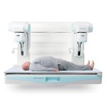
When it comes to your patient’s health, certainty matters. Breast biopsies play a vital role in the continuum of care for women, providing insights into mass and calcification identification so you can make the best-informed decisions.

Dunlee's liquid metal bearing CT replacement tubes extend reach beyond USA and Europe with full registration in Canada and selected Middle Eastern countries.
Groundbreaking technologies have improved our ability to accurately diagnose breast cancer and reduce callbacks, focusing on compassionate care for patients as they move along their breast health journey.
“With Your Stories – lifetime healthcare support” is the future-driven approach to benefit patients through even better prevention, diagnosis, treatment and follow-up and thus help them in the pursuit of a healthy life.
Conventional CT imaging has reached its technical limitations: Resolution can only be improved by small margins and dose cannot be reduced significantly: Photon-counting technology enables drastic improvements.