News • CT, MRI, X-ray, AI, PACS, endoscopy, ultrasound
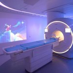
Fujifilm Healthcare acquires Hitachi Diagnostic Imaging, presents new portfolio
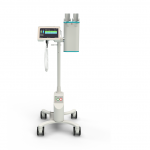
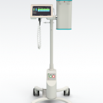
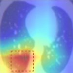
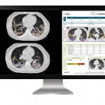
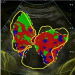
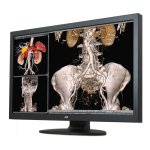
At a virtual European event, Fujifilm Healthcare Europe presented a complete and integrated portfolio of diagnostic products and services, including CT, MRI, X-ray, AI, PACS, endoscopy and ultrasound systems. This launch follows the completion of Fujifilm's acquisition and takeover of Hitachi's Diagnostic Imaging-related business on 31 March 2021 for 179 billion yen (€1.3 billion).