Image source: Unsplash/National Cancer Institute
News • Better tissue discrimination, lower radioation dose
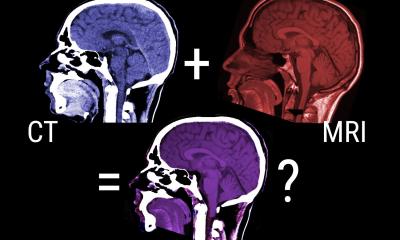
Improving image quality of CT scans
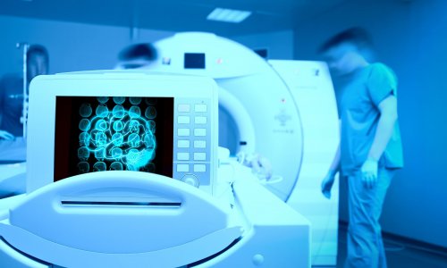
Computed tomography (CT) is one of the most effective medical tests for analysing the effects of many illnesses, including COVID-19, on the lungs. An international team led by the Universitat Oberta de Catalunya (UOC) has developed a new method that improves the quality of the images obtained from CT scans.
The algorithm, which has been tested on simulated data, enables them to distinguish different body’s tissue types better and opens the door to lowering the doses of radiation to which patients are exposed during this type of test.
The researchers published their findings in the Journal of Information Processing.
Our viewpoint was proposing a post-processing approach that does not need a substantial hardware reconfiguration and gives more freedom to imaging scientists for further exploration
Mohammad Mahdi Dehshibi
To reduce the radiation a patient is exposed to during a CT scan, the team led by Mohammad Mahdi Dehshibi, a postdoctoral researcher at the UOC's Scene Understanding and Artificial Intelligence laboratory (SUNAI), of the UOC's Faculty of Computer Science, Multimedia and Telecommunications, of the Pattern Research Centre in Teheran, Iran, has developed a new post-processing algorithm which increases the quality of reconstructed CT images. While conventional CT methods pick up only a part of the X-ray energy spectrum, the researchers tested a broader energy range, divided into intervals, to reach higher contrast. After testing it on constructed data using GATE/GEANT4 simulation software, they found that the algorithm enhances the quality of the images while reducing noise, which enables better discrimination between different types of tissue with lower doses of X-rays.
Recommended article
Interview • Chest X-ray, CT and more
Imaging the coronavirus disease COVID-19
Chest X-ray is the first imaging method to diagnose COVID-19 coronavirus infection in Spain, but in the light of new evidence this may change soon, according to Milagros Martí de Gracia, Vice President of the Spanish Society of Radiology (SERAM) and head of the emergency radiology unit at La Paz Hospital in Madrid, one of the hot spots for viral re-production of COVID-19.
"Distinguishing between two different tissues (either normal or abnormal ones) in the same region is critical for physicians or radiologists to plan for further treatments, where this decision is dealing with the patients' lives," he said. "Having better tissue discrimination increases the success rate of the medicine's plan." The new method increases the capacity to distinguish between tissues by 60% in simulations compared to conventional CT. "Our viewpoint was proposing a post-processing approach that does not need a substantial hardware reconfiguration and gives more freedom to imaging scientists for further exploration," Dehshibi said. "We hope that the findings of this study are later examined in the clinical setting to reduce the radioactive effect of irradiating with X-ray."
Source: Universitat Oberta de Catalunya (UOC)
12.05.2020