News • Diagnosing Pneumonia
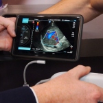
Is lung ultrasound a safe substitute for chest X-Ray?
Lung ultrasound has been shown to be highly effective and safe for diagnosing pneumonia in children and a potential substitute for chest X-ray, according to a study conducted at the Icahn School of Medicine at Mount Sinai.