Article • Preparing for the unpredictable
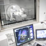
The role of radiology in mass casualty incidents
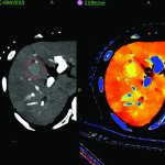
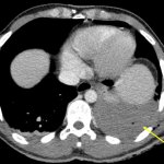
CT has a critical role to play in management of mass casualty incidents with the ability to image patients from head to toe, offering a rapid overview for clinicians. The benefits of CT were outlined by Dr Elizabeth Dick during an ECR session examining the role of radiology in the management of mass casualty incidents, terror attacks and assaults.