News • Biomarker
A real-time MRI method for the early detection of stroke
An international research group at the Max Planck Institute for Biological Cybernetics developed a brain imaging method for improved and early assessment of stroke. Their study presents a methodology which monitors calcium ion fluctuations in the brain using a molecular functional MRI approach.
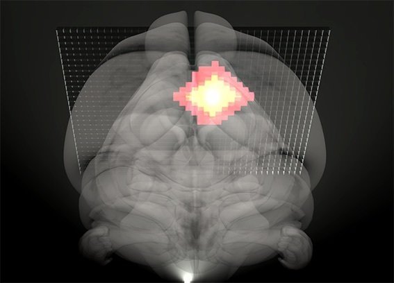
The researchers reported observations from high-resolution imaging of the changing amount of calcium in the brain tissue during stroke. Their reported novel method shows precisely the onset and course of ischemic stroke, the severity of which can be investigated by a three-dimensional MR image reconstruction. Specifically, they used a calcium-sensitive contrast agent as a biomarker, which enabled the changes in calcium concentration to be traced and thus correlated with the spatiotemporal dynamics of cerebral ischemia.
“The commonly used imaging techniques for ischemia diagnosis, such as ultrasonography or computed tomography, often lack the necessary sensitivity to detect ischemia at an early stage,” comments Goran Angelovski, the principal investigator of this study. “We consider our method an important step forward in developing reliable and precise neuroimaging techniques for non-invasive investigations of brain function and dysfunction. It is highly specific for calcium, a messenger of various cellular signals, allowing better differentiation of damaged regions at an early stage of brain injury.”
Cerebral ischemia is characterized in conditions of a reduced blood supply to the brain tissue, causing a deprivation of oxygen and glucose delivery and a production failure of adenosine triphosphate (ATP), a key energy carrier. The resulting energy crisis can trigger a cascade of detrimental biochemical and physiological events, consequently leading to acute or delayed cell death.
Revealing immediate changes during reperfusion are essential for the appropriate treatment and subsequent recovery of brain tissues. Given that the physiological changes in damaged brain tissue occur rather fast, the method may prove ideal for the prompt detection of ischemic onset, and can offer substantial insights into the neurobiology of various neurological disorders.
Source: Max Planck Institute for Biological Cybernetics
26.09.2019