

News • Thyroid cancer and meningioma
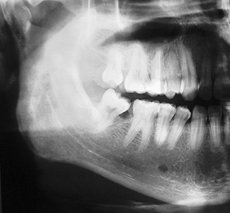
Dental X-rays may increase cancer risk
Research by team at Brighton and Sussex Medical School (BSMS) showed that repeated exposures to dental X-rays may be associated with an increased risk of thyroid cancer and meningioma.
About 3,500 new cases of thyroid cancer and 1,850 cases of meningiomas are diagnosed each year in the UK, and the incidence of both cancers has increased in many countries during the past three decades. For thyroid cancer, much of this increase is probably due to increased surveillance, screening and over-diagnosis (i.e. detection of a cancer that would not ultimately cause symptoms), but the researchers believe other causes need investigation as well.
They published their findings in the journal Thyroid.
The thyroid gland is situated in the neck and the meninges cover the brain and spinal cord – these organs will be exposed to radiation from dental X-rays. Both organs are highly radiosensitive, particularly in childhood and adolescence. Dental radiography, a source of low-dose diagnostic radiation, is often overlooked as a potential hazard to these organs.
The researchers conducted a systematic review and meta-analysis, which summarised the findings from all the previously published studies on dental X-rays exposure and the risk of thyroid cancer, meningioma and other cancers of the head and neck region. They said the results of their research should be treated with caution because these studies did not include individual organ doses and ages at exposure, and are subject to recall bias and other limitations. The researchers said that their synthesis provides good evidence to warrant more research based on dental X-rays records and patient follow-up to test the hypothesis further.
These findings manifest the need to reduce diagnostic radiation exposure as much as possible
Anjum Memon
Professor Memon said: “Little is known about the impact and magnitude of risk associated with dental X-rays, which have been the fastest growing source of human exposure to low-dose ionizing radiation during the past three decades – with many patients being exposed to dental X-rays on multiple occasions over many years. Given this high life-time prevalence and frequency of exposure, even a small associated increase in cancer risk would be of considerable public health importance.”
He said: “The clinical and public health implications of these findings are relevant in light of the increasing incidence of thyroid cancer and meningioma in many countries. Our study highlights the concern that like chest (or other upper body) X-rays, dental X-rays should be prescribed when the patient has a specific clinical need and not as a standard part of evaluation for new patients, for routine check-up, or for periodic screening for dental caries/decay in children/adolescents. Current UK, European and USA guidelines stress the need for thyroid shielding during dental radiography. The findings also stress the need for maintaining comprehensive long-term dental X-rays records, which could follow the patient when they register with a new dentist – thus avoiding the need for unnecessary X-rays.”
Recommended article

Article • 'Is it safe?'
Effective communication on radiation risks
Communicating radiation risks is not only a legal requirement, it is also a moral obligation, asserts Dr Shane J Foley, radiographer and assistant professor at the UCD School of Medicine in Dublin, Ireland. Passing on radiation information has its pitfalls, but several helpful tools can improve communication, some of which the expert highlighted during ECR 2018.
He concluded: “The notion that low-dose radiation exposure through dental radiography is completely without risk needs to be investigated further. Although the individual risk, with modern technology and equipment is likely to be very low, the proportion of the population exposed is high. Considering that about one-third of the general population in developed countries is routinely exposed to one or more dental X-rays per year, these findings manifest the need to reduce diagnostic radiation exposure as much as possible.”
The research team of Professor Anjum Memon with Dr Imogen Rogers, Dr Priya Paudyal and Dr Josefin Sundin called for further studies based on dental X-rays records and patient follow-up.
Source: Brighton and Sussex Medical School (BSMS)
29.10.2019
