Article • Radioembolization
Breast cancer metastasis: benefits and limitations of trans-arterial therapy
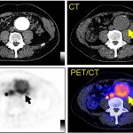
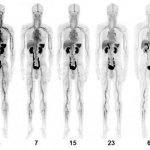
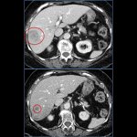
Evidence that radioembolization, a trans-arterial therapy, is safe and stops disease progression in metastatic breast cancer is increasing, a prominent American interventional radiologist showed at the Spectrum conference in Miami.