News • Mobile point-of-care imaging
An MRI-equipped ambulance: A game-changer for stroke care?
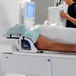
Because time is brain: To explore the potential for accelerated stroke diagnostics, US researchers equipped an ambulance with a portable MRI – with promising first results.