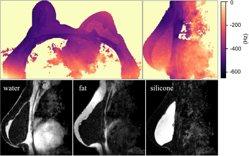
Image source: Stelter et al., IEEE Transactions on Medical Imaging 2022 (CC BY 4.0)
News • Separation between silicone and fat tissue
Algorithm improves MRI breast implant monitoring
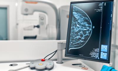
Magnetic resonance imaging (MRI) is a sensitive method for monitoring silicone implants. However, reliable implant examination can be challenging due to the difficulties of separating silicone and fat tissue in the images.
An interdisciplinary research team at the Technical University of Munich (TUM) has now developed a new algorithm that improves the quality of MR images by depicting water, fat, and silicone simultaneously in a reliable and fully automated way.
The researchers published their findings in the journal IEEE Transactions on Medical Imaging.
Silicone implants are commonly used for breast reconstruction after mastectomy and for breast augmentation. Examining these implants at regular intervals helps to detect complications like implant rupture or implant associated anaplastic large cell lymphoma (BI-ALCL) early. MRI is the most sensitive available method for monitoring silicone implants. However, until now, it has been challenging to separate fat and silicone in MRI scans, as these materials generate a similar signal in MRI due to their similar frequencies under the main magnetic field of the MR scanner.
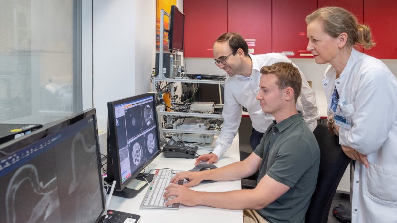
© Andreas Heddergott / TUM
A research team led by Dimitrios Karampinos, Professor of Experimental Magnetic Resonance Imaging at TUM, has now developed a novel processing algorithm to solve the problem of reliably separating water, fat and silicone in MR images. It is based on a specialized data acquisition scheme encoding multiple chemical species previously proposed by his team. “From a technical viewpoint, it is challenging to obtain reliable information about water, fat, and silicone simultaneously with one scan. By collaborating closely with the radiologists at TUM, we not only solved this complex optimization problem from a mathematical perspective, but we developed a solution that can easily be implemented in common clinical MRI scanners and integrated in a clinical workflow”, says Prof. Karampinos.
The imaging process with the new method is simple and the data processing is completely automated. It is intended for breast imaging in all patients, with and without implants. The algorithm uses three main principles to solve the complex problem:
- Hierarchy: The algorithm processes the output from the MRI along a series of steps to create high-quality water, fat and silicone images. At each step, the algorithm decides how to proceed based on the available information, such as for example “implant or no implant”.
- Multi-resolution: High-resolution images depict a lot of detail, which can be helpful for diagnosis. However, high-resolution images are more susceptible to noise and could therefore make it difficult to separate water, fat and silicone. High-resolution images also need considerably more processing time. Hence, the algorithm starts with low-resolution images and increases resolution and complexity along the different steps when needed.
- Graph-cuts: To solve the complex problem of differentiating between several different chemical species, every value of the 3D MRI image is encoded in a corresponding graph. However, one graph alone does not provide enough information to decide reliably if this value corresponds to fat, water or silicone. This is where the algorithm comes into play: Graphs are solved sequentially to build up on the previous information and to find the optimal solution – water, fat or silicone – that makes the most sense with respect to the constraints of the graph and within the context of the whole 3D image.
So far, the new algorithm has shown reliable results even for different types of implants. What is more, acquiring all the information at once instead of one species after the other reduces total scanning time
Eva Maria Fallenberg
Previous methods for MRI examinations are based on suppressing the other materials while imaging the material in question, for example silicone. However, this technique relies on several manual calibration steps, which can be prone to errors. While ruptures of implants can be imaged well with these available methods, slighter changes such as gel bleeding are more difficult to detect.
“The new method is fully automated and does not need any previous calibration and training for the operator. This makes it more robust and reliable than techniques that depend on the suppression of selected materials”, says Dr. Eva Maria Fallenberg, Adjunct Teaching Professor and senior physician at the Institute for diagnostic and interventional radiology at the University hospital Klinikum rechts der Isar. “So far, the new algorithm has shown reliable results even for different types of implants. What is more, acquiring all the information at once instead of one species after the other reduces total scanning time. This improves patient comfort, and allows us to examine more patients.”
After the first promising results, the new imaging process is now being evaluated with a larger cohort of people on clinical MRI scanners commonly used in hospitals. No extra equipment is necessary to adapt the new method. Hence, once the new method has been proven successful in a large patient cohort, an implementation in the large routine clinical setting might be expected. In the longer term, the researchers are also evaluating if the new method provides advantages for assessing non-implant tissue, as it might deliver additional useful information on measuring breast density and depicting breast calcifications.
Source: Technical University of Munich
18.09.2022