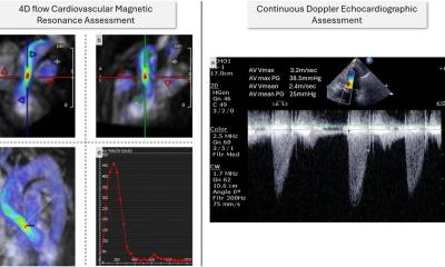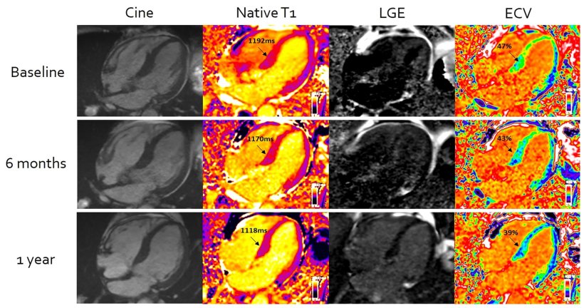
Image source: Martinez-Naharro et al., European Heart Journal 2022 (CC BY 4.0)
News • CMR-ECV vs. light-chain cardiac amyloidosis
'Stiff heart syndrome': Advanced MRI benefits patients
An advanced form of cardiac MRI, developed by academics at University College London (UCL) in collaboration with the Royal Free Hospital, has for the first-time enabled clinicians to measure the effectiveness of chemotherapy in patients with the life-limiting condition ‘stiff heart syndrome’.
Researchers say the breakthrough, published in the European Heart Journal, means doctors will now be able to better guide treatment strategies and, by doing so, improve patients’ prognosis.
Light-chain cardiac amyloidosis (stiff heart syndrome) occurs when plaques of protein called amyloid build up in heart muscle, affecting its ability to pump blood, and without treatment can rapidly lead to heart failure and death. However, assessing the condition has been difficult, as while clinicians can detect the presence of amyloid in the heart, there has been no safe test to measure the amount. This has also meant there has been no way of measuring the therapeutic effect of chemotherapy – the normal first line treatment.
Knowing the amount, rather than just the presence of amyloid, means they can better guide treatment option, by more accurately deciding timing and protocol of second line chemotherapy treatments
Ana Martinez-Naharro
A patient’s response is currently assessed with indirect biological markers, but these do not measure the amount (or reduction) of cardiac amyloid – the drug’s ultimate target – and doctors find the markers less useful when trying to assess second-line chemotherapy treatments. To overcome this, UCL researchers at the National Amyloidosis Centre, have, for 10 years, been developing and refining Cardiovascular Magnetic Resonance (CMR) Extracellular Volume Mapping (ECV) for amyloid. This non-invasive technique enables clinicians to measure both the presence and amount of amyloid protein using MRI. Now for the first time they have used the technology to evaluate the success of chemotherapy treatment, by assessing cardiac amyloid regression or progression.
For the study 176 patients with light-chain cardiac amyloidosis had Cardiovascular Magnetic Resonance with Extracellular Volume Mapping. CMR scans with ECV mapping were done at diagnosis, and then at six, 12 and 24 months after starting chemotherapy. The advanced MRI technique allowed researchers to accurately measure the amount of amyloid protein in hearts, and, for the first-time ever, to measure the changes in response to chemotherapy on repeat scans. By measuring the changes they could detect which patients would have a better or worse prognosis. Further, combining the results with blood tests for the disease, it was found almost 40% of patients had a substantial improvement (reduction) in amyloid deposition, something that was not thought to be possible before – showing how effective chemotherapy can be.
First author, Dr Ana Martinez-Naharro (UCL Division of Medicine) said: “In this study we were assessing the ability and value of CMR to carry out ECV mapping to measure directly the changes in amyloid proteins in the heart in response to chemotherapy and how they correlate with the indirect markers that current exist. The scans and data made available using this technique, gave us the information to both see the amount of amyloid protein and also the regression in amyloid during the course of chemotherapy treatment. This is incredibly valuable for clinicians; knowing the amount, rather than just the presence of amyloid, means they can better guide treatment option, by more accurately deciding timing and protocol of second line chemotherapy treatments.”
Recommended article

Article • CAD pros and cons
CT or MRI – which is better for imaging stable cardiac chest pain?
When it comes to imaging stable cardiac chest pain, which modality should be used as the first-line test to investigate coronary artery disease: CT or MRI? Radiologists discussed the strengths and limitations of the two approaches in a lively Pros and Cons session at ECR Overture.
In the UK, there are about 4,000 to 6,000 amyloidosis patients, however there are many more who remain undiagnosed. The condition is more prevalent in men than women.
Senior author, Professor Marianna Fontana (UCL Division of Medicine), a British Heart Foundation (BHF) Clinical Fellow, said this UCL-developed MRI technique should now be used immediately to diagnose and assess all cases of light-chain cardiac amyloidosis. “Since MRI scans are widely available, by developing the use of ECV mapping in a machine that already is used for these patients, we hope that its use can be made available to more patients to help improve their care. The aim would be to use these scans routinely for all patients with the disease to help doctors monitor the response to chemotherapy to help improve patient survival, which is very poor in patients who do not respond to treatment.”
For the single-centre trial, patients were recruited and treated at the National Amyloidosis Centre, which is part of the UCL Division of Medicine and based at the Royal Free Hospital, London. Professor Fontana added: “Twenty years ago, another MRI technique called T2*, was proven to be able to do the same thing for measuring the amount of iron in the heart in a condition called thalassemia. For the last two decades, it has been used to help monitor patients’ response to treatment and allow doctors to make changes along the way. As a result, survival has improved for thalassemia. As T2* has changed the landscape for this condition, tracking changes using ECV mapping of the heart may completely change the landscape for patients with amyloidosis, with the potential of guide treatment and, in the process, lead to improvements in patient outcomes.”
Source: University College London
29.07.2022